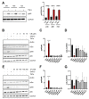
Perturbation of the Actin Cytoskeleton in Human Hepatoma Cells Influences Interleukin-6 (IL-6) Signaling, but Not Soluble IL-6 Receptor Generation or NF-κB Activation
- PMID: 34281231
- PMCID: PMC8268250
- DOI: 10.3390/ijms22137171
Perturbation of the Actin Cytoskeleton in Human Hepatoma Cells Influences Interleukin-6 (IL-6) Signaling, but Not Soluble IL-6 Receptor Generation or NF-κB Activation
Abstract
The transcription factor nuclear factor-kappa B (NF-κB) is critically involved in inflammation and cancer development. Activation of NF-κB induces the expression and release of several pro-inflammatory proteins, which include the cytokine interleukin-6 (IL-6). Perturbation of the actin cytoskeleton has been previously shown to activate NF-κB signaling. In this study, we analyze the influence of different compounds that modulate the actin cytoskeleton on NF-κB activation, IL-6 signaling and the proteolytic generation of the soluble IL-6 receptor (sIL-6R) in human hepatoma cells. We show that perturbation of the actin cytoskeleton is not sufficient to induce NF-κB activation and IL-6 secretion. However, perturbation of the actin cytoskeleton reduces IL-6-induced activation of the transcription factor STAT3 in Hep3B cells. In contrast, IL-6R proteolysis by the metalloprotease ADAM10 did not depend upon the integrity of the actin cytoskeleton. In summary, we uncover a previously unknown function of the actin cytoskeleton in IL-6-mediated signal transduction in Hep3B cells.
Keywords: NF-κB; STAT3; actin; interleukin-6; interleukin-6 receptor.
Conflict of interest statement
The authors declare no conflict of interest. The funders had no role in the design of the study; in the collection, analyses, or interpretation of data; in the writing of the manuscript, or in the decision to publish the results.
Figures


Similar articles
-
Persistent activation of signal transducer and activator of transcription 3 via interleukin-6 trans-signaling is involved in fibrosis of endometriosis.Hum Reprod. 2022 Jun 30;37(7):1489-1504. doi: 10.1093/humrep/deac098. Hum Reprod. 2022. PMID: 35551394
-
Melittin enhances apoptosis through suppression of IL-6/sIL-6R complex-induced NF-κB and STAT3 activation and Bcl-2 expression for human fibroblast-like synoviocytes in rheumatoid arthritis.Joint Bone Spine. 2011 Oct;78(5):471-7. doi: 10.1016/j.jbspin.2011.01.004. Epub 2011 Feb 26. Joint Bone Spine. 2011. PMID: 21354845
-
Activation of Toll-like Receptor 2 (TLR2) induces Interleukin-6 trans-signaling.Sci Rep. 2019 May 13;9(1):7306. doi: 10.1038/s41598-019-43617-5. Sci Rep. 2019. PMID: 31086276 Free PMC article.
-
Hepatic acute phase proteins--regulation by IL-6- and IL-1-type cytokines involving STAT3 and its crosstalk with NF-κB-dependent signaling.Eur J Cell Biol. 2012 Jun-Jul;91(6-7):496-505. doi: 10.1016/j.ejcb.2011.09.008. Epub 2011 Nov 16. Eur J Cell Biol. 2012. PMID: 22093287 Review.
-
Role of interleukin-6 in cachexia: therapeutic implications.Curr Opin Support Palliat Care. 2014 Dec;8(4):321-7. doi: 10.1097/SPC.0000000000000091. Curr Opin Support Palliat Care. 2014. PMID: 25319274 Free PMC article. Review.
Cited by
-
Aggravated Ulcerative Colitis via circNlgn-Mediated Suppression of Nuclear Actin Polymerization.Research (Wash D C). 2024 Aug 23;7:0441. doi: 10.34133/research.0441. eCollection 2024. Research (Wash D C). 2024. PMID: 39183944 Free PMC article.
-
Extracellular Ribosomal RNA Acts Synergistically with Toll-like Receptor 2 Agonists to Promote Inflammation.Cells. 2022 Apr 24;11(9):1440. doi: 10.3390/cells11091440. Cells. 2022. PMID: 35563745 Free PMC article.
References
MeSH terms
Substances
Grants and funding
LinkOut - more resources
Full Text Sources
Research Materials
Miscellaneous

