. 2021 Jul 3;22(13):7189.
doi: 10.3390/ijms22137189.

Bisphenol A Modulates Autophagy and Exacerbates Chronic Kidney Damage in Mice

Affiliations

Bisphenol A Modulates Autophagy and Exacerbates Chronic Kidney Damage in Mice

Alberto Ruiz Priego et al. Int J Mol Sci. .

Abstract

Background: Bisphenol A (BPA) is a ubiquitous environmental toxin that accumulates in chronic kidney disease (CKD). Our aim was to explore the effect of chronic exposition of BPA in healthy and injured kidney investigating potential mechanisms involved.

Methods: In C57Bl/6 mice, administration of BPA (120 mg/kg/day, i.p for 5 days/week) was done for 2 and 5 weeks. To study BPA effect on CKD, a model of subtotal nephrectomy (SNX) combined with BPA administration for 5 weeks was employed. In vitro studies were done in human proximal tubular epithelial cells (HK-2 line).

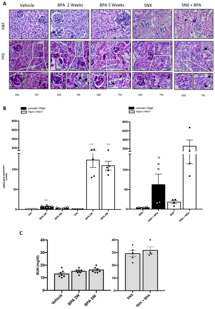
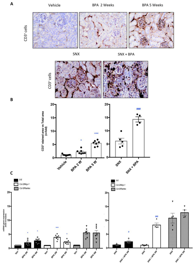
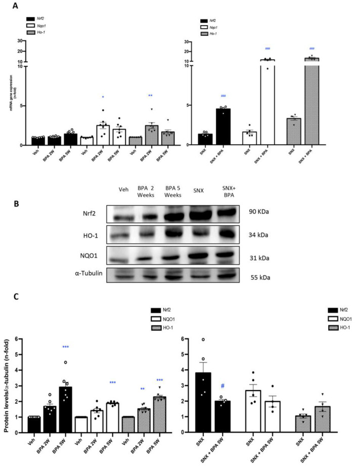
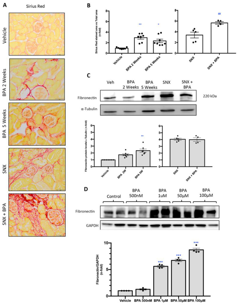
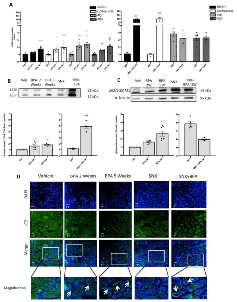
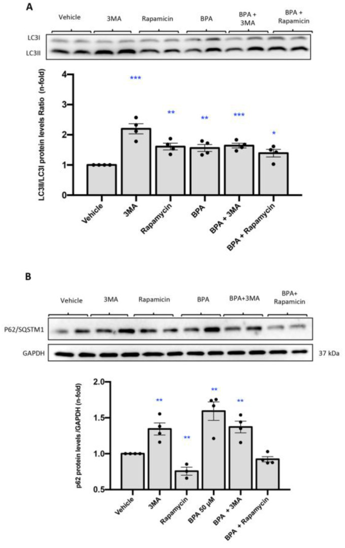
Results: Chronic BPA administration to healthy mice induces inflammatory infiltration in the kidney, tubular injury and renal fibrosis (assessed by increased collagen deposition). Moreover, in SNX mice BPA exposure exacerbates renal lesions, including overexpression of the tubular damage biomarker Hepatitis A virus cellular receptor 1 (Havcr-1/KIM-1). BPA upregulated several proinflammatory genes and increased the antioxidant response [Nuclear factor erythroid 2-related factor 2 (Nrf2), Heme Oxygenase-1 (Ho-1) and NAD(P)H dehydrogenase quinone 1 (Nqo-1)] both in healthy and SNX mice. The autophagy process was modulated by BPA, through elevated autophagy-related gene 5 (Atg5), autophagy-related gene 7 (Atg7), Microtubule-associated proteins 1A/1B light chain 3B (Map1lc3b/Lc3b) and Beclin-1 gene levels and blockaded the autophagosome maturation and flux (p62 levels). This autophagy deregulation was confirmed in vitro.

Conclusions: BPA deregulates autophagy flux and redox protective mechanisms, suggesting a potential mechanism of BPA deleterious effects in the kidney.

Keywords: Bisphenol A; autophagy; fibrosis; inflammation; oxidative stress.


Conflict of interest statement

The authors declare no conflict of interest.

Figures

Figure 1
Figure 1
Systemic chronic administration of BPA induces kidney damage in healthy mice and exacerbates renal lesions in an experimental model of progressive CKD. Animals were intraperitoneal (i.p) injected with 120 mg/kg/day BPA (5 days a week) or vehicle (corn oil) and sacrificed after 2 or 5 weeks. Some animals were subjected to subtotal nephrectomy (SNX), then treated or not with BPA and sacrificed after 5 weeks. (A) Representative pictures of H&E and PAS staining show tubular dilatation, mesangial matrix proliferation, glomerulus disorganization and loss of glomerular/tubular basement membrane and the brush border of the proximal tubules in animals exposed to BPA mainly in SNX compared to vehicle mice. Magnification 200× (GM: glomerular magnification; TM: Tubular magnification; arrows indicate tubular damage /scale bar appears in lower right area of the image and represents 50 µm). (B) Renal mRNA expression of Havcr-1/Kim-1 and Lipocalin2 (that codify KIM-1 and N-GAL, respectively) were evaluated by RT-PCR. (C) Serum Urea Nitrogen (BUN) levels are shown in mg/dl. All data are presented as the mean ± SEM of 4–7 mice per group; *** p < 0.001 vs. Vehicle, # p < 0.05 vs. SNX. Comparison between SNX and SNX + BPA groups was performed using a parametric unpaired two-sided t test. The analysis of WT vs. BPA-treated groups was done by one-way ANOVA, followed by the Tukey HSD multiple comparison Test.
Figure 2
Figure 2
Systemic chronic administration of BPA causes interstitial inflammatory cell infiltration in the kidney and increases renal expression of proinflammatory factors. Animals were intraperitoneal injected with 120 mg/kg/day BPA (5 days a week) or vehicle (corn oil) and sacrificed after 2 or 5 weeks. Some animals were subjected to subtotal nephrectomy (SNX), then treated or not with BPA and sacrificed after 5 weeks. (A) Paraffin-embedded kidney sections were stained with an anti-CD3+ antibody. Representative immunohistochemistry pictures identifying inflammatory T cell infiltration (CD3+ T lymphocytes). Magnification 200× (scale bar appears in lower right area of the image and represents 50 µm). (B) Immunohistochemistry staining quantification expressed as mean of stained area vs. total area ± SEM of 4–7 animals per group. (C) Gene expression of Il-6, Ccl-2 (Mcp-1) and Ccl-5 (Rantes) were evaluated by RT-PCR. Data are expressed as mean ± SEM of 4–7 animals per group. * p < 0.05; *** p < 0.001 vs. Vehicle, # p < 0.05; ### p < 0.001 vs. SNX. Comparison between SNX and SNX + BPA groups was performed using a parametric unpaired two-sided t test. The analysis of WT vs. BPA-treated groups was done by one-way ANOVA, followed by the Tukey HSD multiple comparison test.
Figure 3
Figure 3
Systemic chronic administration of BPA activates Nrf2 pathway in SNX model of renal damage. Animals were intraperitoneal injected with 120 mg/kg/day BPA (5 days a week) or vehicle (corn oil) and sacrificed after 2 or 5 weeks. Some animals were subjected to subtotal nephrectomy (SNX), then treated or not with BPA and sacrificed after 5 weeks. (A) mRNA expression of Nrf2 and Nrf2-regulated genes: Nqo-1 Ho-1 evaluated by RT-PCR in renal tissue. (B,C) Representative Western blot image and the quantification of Nrf2, NQO1 and HO-1 levels in renal tissue lysates. All data are presented as the mean ± SEM of 4–7 mice per group, * p < 0.05; ** p < 0.01; *** p < 0.001 vs. Vehicle, # p < 0.05; ### p < 0.001 vs. SNX. Comparison between SNX and SNX + BPA groups was performed using a parametric unpaired two-sided t test. The analysis of WT vs. BPA-treated groups was done by one-way ANOVA, followed by the Tukey HSD multiple comparison Test.
Figure 4
Figure 4
Systemic chronic administration of BPA induces renal fibrosis in mice. Animals were intraperitoneal injected with 120 mg/kg/day BPA (5 days a week) or vehicle (corn oil) and sacrificed after 2 or 5 weeks. Some animals were subjected to subtotal nephrectomy (SNX), then treated or not with BPA and sacrificed after 5 weeks. (A,B) Collagen deposition was evaluated in paraffin-embedded sections by Sirius Red staining; quantification assessed the stained area vs. total area. (A) Figures show a representative picture from each group. Magnification 200×. (scale bar appears in lower right area of the image and represents 50 µm). (B) The quantification of Sirius Red staining. (C) Fibronectin protein levels were evaluated in total renal extracts by Western blot. Figures shows representative mice from each group and the quantification of the Western blot data. Data are expressed as mean ± SEM of 4–7 animals per group. * p < 0.05 vs. control; *** p < 0.001 vs. control; ## p < 0.01 vs. SNX. (D) Treatment with BPA increases ECM proteins production in cultured renal cells. HK2 cells were treated with BPA at dose of 500 nM, 1, 50 and 100 μM for 48 h. Fibronectin levels were detected by Western blot. Figures shows representative experiment and the quantification of the Western blot. All data are presented as the mean ± SEM of 4 experiments, * p < 0.05; ** p < 0.01; *** p < 0.001 vs. Vehicle, ## p < 0.01; vs. BPA. Comparison between SNX vs. SNX + BPA groups was performed using a parametric unpaired two-sided t test. The analysis of WT vs. BPA-treated groups was done by one-way ANOVA, followed by the Tukey HSD multiple comparison Test.
Figure 5
Figure 5
Systemic chronic administration of BPA blocks autophagosome maturation in experimental SNX model. Animals were intraperitoneal injected with 120 mg/kg/day BPA (5 days a week) or vehicle (corn oil) and sacrificed after 2 or 5 weeks. Some animals were subjected to subtotal nephrectomy (SNX), then treated or not with BPA and sacrificed after 5 weeks. (A) Beclin-1, Atg5, Atg7, Map1lc3b/Lc3b mRNA levels were assessed by RT-PCR in renal tissue. (B,C) Representative Western blot and quantification of protein levels of LC3II/I ratio and p62/SQSTM1 in renal tissue lysates. All data are presented as the mean ± SEM of 4–7 mice per group, ** p < 0.01; *** p < 0.001 vs. Vehicle, ### p < 0.001 vs. SNX. (D) Immunofluorescence staining localized LC3 expression in renal tubules in all groups with exposure to BPA. Magnification 200×; scale bar represents 20 µm (arrows identify LC3B cytoplasmic staining). Comparison between SNX and SNX + BPA groups was performed using a parametric unpaired two-sided t test. The analysis of WT vs. BPA-treated groups was done by one-way ANOVA, followed by the Tukey HSD multiple comparison Test.
Figure 6
Figure 6
Treatment with BPA causes LC3B accumulation in cultured renal cells compared to 3MA and Rapamycin. (A) HK2 cells were treated with BPA at dose of 50 μM for 24 h. The level of LC3II/I was detected by Western blot. LC3II/I protein levels ratio of. BPA treatment induced p62/SQSTM1 accumulation in HK2 cells compared to 3MA and Rapamycin. (B) HK2 cells were treated with BPA at dose of 50 μM for 24 h. The level of p62/SQSTM1 was detected by Western blot. p62/SQSTM1 protein levels quantification. All data are presented as the mean ± SEM of 4 experiments, * p < 0.05; ** p < 0.01; *** p < 0.001 vs. control.

Similar articles

Cited by

References

    1. González-Parra E., Herrero J.A., Elewa U., Bosch R.J., Arduán A.O., Egido J. Bisphenol A in chronic kidney disease. Int. J. Nephrol. 2013;2013:437857. doi: 10.1155/2013/437857. - DOI - PMC - PubMed
    1. Rubin B.S. Bisphenol A: An endocrine disruptor with widespread exposure and multiple effects. J. Steroid Biochem. Mol. Biol. 2011;127:27–34. doi: 10.1016/j.jsbmb.2011.05.002. - DOI - PubMed
    1. Kang J.H., Kondo F., Katayama Y. Human exposure to bisphenol A. Toxicology. 2006;226:79–89. doi: 10.1016/j.tox.2006.06.009. - DOI - PubMed
    1. Thomson B.M., Grounds P.R. Bisphenol A in canned foods in New Zealand: An exposure assessment. Food Addit. Contam. 2005;22:65–72. doi: 10.1080/02652030400027920. - DOI - PubMed
    1. Wilson N.K., Chuang J.C., Morgan M.K., Lordo R.A., Sheldon L.S. An observational study of the potential exposures of preschool children to pentachlorophenol, bisphenol-A, and nonylphenol at home and daycare. Environ. Res. 2007;103:9–20. doi: 10.1016/j.envres.2006.04.006. - DOI - PubMed

MeSH terms