. 2021 Aug 31;12(4):e0127421.
doi: 10.1128/mBio.01274-21. Epub 2021 Jul 20.

Near-Pan-neutralizing, Plasma Deconvoluted Antibody N49P6 Mimics Host Receptor CD4 in Its Quaternary Interactions with the HIV-1 Envelope Trimer

Affiliations

Near-Pan-neutralizing, Plasma Deconvoluted Antibody N49P6 Mimics Host Receptor CD4 in Its Quaternary Interactions with the HIV-1 Envelope Trimer

William D Tolbert et al. mBio. .

Abstract

The first step in HIV-1 entry is the attachment of the envelope (Env) trimer to target cell CD4. As such, the CD4-binding site (CD4bs) remains one of the few universally accessible sites for antibodies (Abs). We recently described a method of isolating Abs directly from the circulating plasma and described a panel of broadly neutralizing Abs (bnAbs) from an HIV-1 "elite neutralizer" referred to as patient N49 (N49 Ab lineage [M. M. Sajadi, A. Dashti, Z. R. Tehrani, W. D. Tolbert, et al., Cell 173:1783-1795.e14, 2018, https://doi.org/10.1016/j.cell.2018.03.061]). Here, we describe the molecular details of antigen recognition by N49P6, an Ab of the N49 lineage that recapitulates most of the neutralization breadth and potency of the donor's plasma IgG. Our studies done in the context of monomeric and trimeric antigens indicate that N49P6 combines many characteristics of known CD4bs-specific bnAbs with features that are unique to the N49 Ab lineage to achieve its remarkable neutralization breadth. These include the omission of the CD4 Phe43 cavity and dependence instead on interactions with highly conserved gp120 inner domain layer 3. Interestingly, when bound to BG505 SOSIP, N49P6 closely mimics the initial contact of host receptor CD4 to the adjacent promoter of the HIV-1 Env trimer to lock the trimer in the closed conformation. Altogether, N49P6 defines a new class of near-pan-neutralizing, plasma deconvoluted CD4bs Abs that we refer to as the N49P series. The details of the mechanisms of action of this new Ab class pave the way for the next generation of HIV-1 bnAbs that can be used as vaccine components of therapeutics. IMPORTANCE Binding to target cell CD4 is the first crucial step required for HIV-1 infection. Thus, the CD4-binding site (CD4bs) is one of the most accessible sites for antibodies (Abs). However, due to steric constraints, only a few Abs are capable of targeting this site. Here, we show that the exceptional neutralization breadth and potency of N49P6, a near-pan-neutralizing Ab targeting the CD4bs isolated from the plasma of an HIV-1 "elite neutralizer," patient N49, are due to its signature combination of more typical CD4bs Ab-binding characteristics with unique interactions with the highly conserved gp120 inner domain. In addition, we also present a structural analysis of N49P6 in complex with the BG505 SOSIP trimer to show that N49P6 exhibits remarkable breadth in part by mimicking CD4's quaternary interaction with the neighboring gp120 protomer. In its mode of antigen interaction, N49P6 is unique and represents a new class of CD4bs-specific bnAbs.

Keywords: CD4-binding site; HIV; N49P lineage; near-pan-neutralizing; neutralizing antibodies.


Figures

FIG 1
FIG 1
Neutralization profiles of mAbs N49P6 and N49P7. (A) A multiclade, 117-HIV-1-pseudovirus panel was tested against mAbs N49P6 and N49P7. IC50 and IC80 values are color-coded according to their potencies. Both mAbs N49P6 and N49P7 exhibited 100% breadth, with all pseudoviruses having an IC50 of <50 μg/ml. (B) Geometric mean IC50 and IC80 values of mAbs N49P6 and N49P7 against each clade and the whole panel.
FIG 2
FIG 2
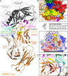
Crystal structure of the N49P6 Fab-gp12093TH057coree complex. (A) Overall structure of the complex shown as a ribbon diagram. The complementarity-determining regions (CDRs) of N49P6 Fab are colored as follows: CDR H1 is black, CDR H2 is yellow, CDR H3 is red, CDR L1 is dark green, and CDR L3 is blue. The outer and inner domains of gp120 are dark and light gray, respectively. The outer domain D and E, CD4 binding, and V5 loops are shown in cyan, purple-blue, magenta, and green, respectively. Carbohydrates at positions N276 (loop D) and N355 (loop E) are shown as sticks. (B) N49P6 Fab-gp12093TH057coree interface with coloring as described above for panel A. (Top) N49P6 Fab is shown as a molecular surface, and the gp120 contact residues are shown as sticks. (Bottom) A 180° view reveals the detailed interaction of N49P6 Fab and the gp120 surface. Contact residues of N49P6 Fab are shown as sticks, and all Fab contact residues are listed. gp120 is shown as a molecular surface and colored according to its electrostatic potential, with red, blue, and white representing negative, positive, and neutral electrostatic potentials, respectively. (C) Blowup view of the hydrogen bond network of the interaction between N49P6 Fab and the gp120 surface. Three hydrogen bonds (CDR H2 W50 and loop D N280, CDR H3 W100J and loop D N279, and CDR L3 E96 and loop D N280) and a salt bridge (heavy chain residue R71 and CD4-binding loop D368) are formed at the interface. Residues contributing to the interaction are shown as sticks, and the hydrogen bond network is shown with red dashed lines.
FIG 3
FIG 3
Structural comparisons of N49P6 Fab- and N49P7-gp12093TH057coree complexes. (A) Blowup view into the Fab-gp120coree interface. The molecular surface is displayed over gp12093TH057coree, and the CDRs of N49P6 are shown as sticks, with colors as described in the Fig. 2 legend. The heavy chain and light chain of N49P7 are shown in dark and light blue, respectively. All structures were superimposed based on gp12093TH057coree. The N49P7 Fab-gp12093TH057coree structure is from PDB accession number 6BCK. (B) Binding footprints for N49P6 and N49P7 on gp12093TH057coree are outlined in yellow. The gp120 surface is colored as described in the Fig. 2 legend. (C) Contact residues of CDRs of N49P6 and N49P7 with gp12093TH057core are mapped onto the primary sequences. Contact residues are defined by a 5-Å cutoff and marked above the sequence with + for the side chain and − for the main chain to indicate the type of contact. Contact types are colored as follows: green for hydrophilic, blue for hydrophobic, and black for both. Buried surface residues were determined by PISA and are shaded in green. Residues that differ between N49P6 and N49P7 are highlighted with dashed boxes. (D) Buried surface area (BSA) contributions to binding of gp120 residues (top) and N49P6/N49P7 Fab residues (bottom) as calculated by PISA. (Top) The gp120 sequence is color-coded in a gradient based on its sequence conservation: dark blue corresponds to the percentage of sequences in the HIV sequence compendium (https://www.hiv.lanl.gov/content/sequence/HIV/COMPENDIUM/compendium.html) that differ at that position from the Hxbc2 reference sequence 0.2 to 7% of the time, and red corresponds those that differ at that position from Hxbc2 87 to 99.9% of the time. Intermediate colors correspond to intermediate percentages on a roughly linear scale. Inner domain layers 2 and 3 and the outer domain D, V5, and CD4-binding loops are colored as described in the legend of Fig. 2. BSA contributions for N49P6 and N49P7 contact residues are shown. (Bottom) CDRs H1, H2, H3, L1, and L3 are colored as described in the legend of Fig. 2. BSA contributions to binding for N49P6 and N49P7 CDR residues are shown in darker and lighter shades of the CDR color, respectively, and those for FWR residues shown in orange and blue, respectively.
FIG 4
FIG 4
Structural comparison of binding modes between N49P6 and other broadly neutralizing CD4-binding-site antibodies. (A) Pie charts showing the buried surface area (BSA) contributions to gp120 binding. The outer domain loops: D, CD4 binding, and V5 are colored as shown. The other outer domain and layers 2 and 3 of inner domain are also colored as shown. (B) Comparison of interactions between layers 2 and 3 of the gp120 inner domain and CDR H3 of N49P6 with other bnAbs (N49P7, VRC01, VRC03, VRC07, NIH45-46, and N6). The molecular surface is displayed over the gp120core, and the CDRs are shown as a ribbon diagram, with coloring as described in the legend of Fig. 2. Layers 2 and 3 of the gp120 inner domain are shown in darker and lighter shades of gray, respectively. Layer 3-contacting residues are labeled on the surface of gp120. (C) BSA contributions to layer 2 and 3 binding for a range of broadly neutralizing CD4bs antibodies (N49P6, N49P7, VRC01, VRC03, VRC07, NIH45-46, and N6). Residues of layers 2 and 3 are color-coded based on their sequence conservation as described in the legend of Fig. 3. BSA values for N49P6, N49P7, VRC01, VRC03, VRC07, NIH45-46, and N6 contact residues are shown. (D) Contact residues of gp120 mapped onto the gp12093TH057core sequences. Contact residues are defined by a 5-Å cutoff and marked above the sequence with + for the side chain and − for the main chain to indicate the type of contact. Buried surface residues were determined by PISA and are shaded blue.
FIG 5
FIG 5
Overall structure of the N49P6 Fab-BG505 SOSIP.664 trimer complex. (A) Side and top views of the crystal structure of a ternary complex of the BG505 SOSIP.664 HIV-1 Env trimer and three N49P6 Fabs (orange) in ribbon representation. Each gp120-gp41 protomer is shown in a different color. The outer domain loops are shown in primary gp120 contact and colored as described in the legend of Fig. 2. (B) Structural comparison of the N49P6 Fab-gp12093TH057coree complex and the N49P6 Fab-gp120 (primary contact) protomer of the BG505 trimer. The complexes are superimposed based on gp12093TH057coree. The N49P6 Fab-gp12093TH057coree complex is colored as described in the legend of Fig. 2, and the heavy and light chains of N49P6 Fab in the N49P6-BG505 protomer complex are shown in dark and lighter shades of blue, respectively. The enlargements show the contacts between N49P6 Fab and gp12093TH057core (top) and the BG505 protomer (bottom). (C) BSA contributions to binding of gp120 (93TH057 core and BG505 gp120 [primary contact] protomer) residues (top) and N49P6 residues (bottom) as calculated by PISA. (Top) The gp120 sequence is color-coded based on its sequence conservation as described in the legend of Fig. 3. Inner domain layers 2 and 3 and the outer domain D, V5, and CD4-binding loops are colored as described in the legend of Fig. 2. BSA values for N49P6 and N49P7 contact residues are shown in blue and red, respectively. (Bottom) All CDRs, H1, H2, H3, L1, and L3, are colored as described in the legend of Fig. 2. BSA contributions to binding of N49P6 contact residues to gp12093TH057coree and the primary BG505 protomer are shown in darker and lighter shades of the CDR color, respectively.
FIG 6
FIG 6
Molecular details of the interprotomer contacts of N49P6. (A) Structural alignment of the N49P6 Fab-BG505 SOSIP.664 trimer complex and the BG505 SOSIP.664 trimer with one CD4 molecule bound (PDB accession number 5U1F). N49P6 Fab (orange) and CD4 (green) are shown as a ribbon diagram. The zoomed-in views depict the contact between N49P6 Fab or CD4 and the adjacent BG505 protomer. All contacting residues are shown as sticks. (B) Networks of interactions formed between N49P6 Fab/CD4 and the adjacent gp120 BG505 protomer as defined by either a 5-Å distance criterion cutoff for N49P6 or a 7.5-Å distance criterion cutoff for CD4 are shown as solid lines. Layer 1 residues are color-coded based on their sequence conservation as described in the legend of Fig. 3. (C) Binding footprints of N49P6 and CD4 on the adjacent gp120 BG505 protomer (surface representation) are colored in yellow. Contacting residues of N49P6 and CD4 are shown in a stick representation (framework region [FWR] in orange, CDR H1 in black, CDR H3 in red, and CD4 in green). (D) Binding footprints of other CD4bs Fabs (VRC01 [PDB accession number 5FYJ], VRC03 [PDB accession number 6CDI], NIH45-46 [PDB accession number 5WDU], 3BNC117 [PDB accession number 5V8M], 1-18 [PDB accession number 6UDJ], and CH31 [PDB accession number 6NNJ]) on the adjacent gp120 protomer (surface representation) are shown as described above for panel C. The contacting residues are defined by a 5-Å distance criterion cutoff. All contact residues are shown in a stick representation, with framework residues in light green and CDR H1 in black.
FIG 7
FIG 7
Structural rearrangements of the HIV-1 Env trimer upon binding to host receptor CD4 and CD4-binding-site-specific bnAbs. (A) Overall structure of the unbound BG505 SOSIP.664 trimer (referred to as the “unbound” BG505 SOSIP conformation) (PDB accession number 4ZMJ) (left) and the trimer with 3 CD4s bound (referred to as the “soluble CD4 [sCD4]-bound” BG505 SOSIP conformation) (PDB accession number 5VN3) (right). Trimers were aligned based on the α-carbon positions of the central gp41 α7 helices, residues 570 to 595, and a center (Centr) (gray sphere) for the alignment calculated from the combined α-carbon positions. The relative position for each gp120 was then calculated based on the α-carbon position for residue 375 at the base of the CD4 Phe43-binding pocket in each gp120 of the trimer (375Cα) (shown as blue spheres). The distances between the center and the 375Cα of each protomer (a, b, and c for “unbound” and a′, b′, and c′ for “sCD4 bound”) and the 375Cα atoms of neighboring protomers (d, e, and f for unbound and d′, e′, and f′ for sCD4 bound) are shown to indicate the extent of the protomer rearrangement after CD4 binding. (B) Comparison of unbound, sCD4-bound, and bnAb-bound conformations of the HIV-1 BG505 SOSIP trimer. The BG505 SOSIP.664 complex structures (N49P6 Fab-BG505 SOSIP.664, VRC01 scFv-x1193.c1 SOSIP.664 [PDB accession number 5FYJ], VRC03 scFv-BG505 SOSIP.664 [accession number 6CDI], NIH45-46 scFv–BG505 SOSIP.664 [accession number 5WDU], 3BNC117 scFv-BG505 SOSIP.664 [accession number 5V8M], CH31 scFv-BG505 SOSIP.664 [accession number 6NNJ], 1-18 scFv–BG505 SOSIP.664 [accession number 6UDJ], and one CD4-bound BG505 SOSIP.664 [accession number 5U1F]) were aligned as described above for panel A, and the 375Cα atom of each complex is shown as colored spheres. Distances for sCD4-bound and unbound BG505 SOSIP trimers are labeled as described above for panel A, with the equivalent N49P6 distances added as red dashes. The clockwise rotation for each gp120 in the sCD4-bound trimer relative to the equivalent gp120 in the closed unbound trimer is also shown and labeled a-a′, b-b′, and c-c′. (C) a, b, c, d, e, and f distances and a-a′, b-b′, and c-c′ rotation angles for each bnAb and CD4 complex relative to the unbound BG505 SOSIP trimer as shown in panel B.

References

    1. Stefic K, Bouvin-Pley M, Braibant M, Barin F. 2019. Impact of HIV-1 diversity on its sensitivity to neutralization. Vaccines (Basel) 7:74. doi:10.3390/vaccines7030074. - DOI - PMC - PubMed
    1. Wu X, Yang ZY, Li Y, Hogerkorp CM, Schief WR, Seaman MS, Zhou T, Schmidt SD, Wu L, Xu L, Longo NS, McKee K, O’Dell S, Louder MK, Wycuff DL, Feng Y, Nason M, Doria-Rose N, Connors M, Kwong PD, Roederer M, Wyatt RT, Nabel GJ, Mascola JR. 2010. Rational design of envelope identifies broadly neutralizing human monoclonal antibodies to HIV-1. Science 329:856–861. doi:10.1126/science.1187659. - DOI - PMC - PubMed
    1. Trkola A, Pomales AB, Yuan H, Korber B, Maddon PJ, Allaway GP, Katinger H, Barbas CF, III, Burton DR, Ho DD, Moore JP. 1995. Cross-clade neutralization of primary isolates of human immunodeficiency virus type 1 by human monoclonal antibodies and tetrameric CD4-IgG. J Virol 69:6609–6617. doi:10.1128/JVI.69.11.6609-6617.1995. - DOI - PMC - PubMed
    1. Gray ES, Madiga MC, Moore PL, Mlisana K, Abdool Karim SS, Binley JM, Shaw GM, Mascola JR, Morris L. 2009. Broad neutralization of human immunodeficiency virus type 1 mediated by plasma antibodies against the gp41 membrane proximal external region. J Virol 83:11265–11274. doi:10.1128/JVI.01359-09. - DOI - PMC - PubMed
    1. Huang J, Kang BH, Pancera M, Lee JH, Tong T, Feng Y, Imamichi H, Georgiev IS, Chuang GY, Druz A, Doria-Rose NA, Laub L, Sliepen K, van Gils MJ, de la Pena AT, Derking R, Klasse PJ, Migueles SA, Bailer RT, Alam M, Pugach P, Haynes BF, Wyatt RT, Sanders RW, Binley JM, Ward AB, Mascola JR, Kwong PD, Connors M. 2014. Broad and potent HIV-1 neutralization by a human antibody that binds the gp41-gp120 interface. Nature 515:138–142. doi:10.1038/nature13601. - DOI - PMC - PubMed

Publication types

MeSH terms