. 2021 Jul 19;19(1):218.
doi: 10.1186/s12951-021-00960-y.

Growth rate-dependent flexural rigidity of microtubules influences pattern formation in collective motion

Affiliations

Growth rate-dependent flexural rigidity of microtubules influences pattern formation in collective motion

Hang Zhou et al. J Nanobiotechnology. .

Abstract

Background: Microtubules (MTs) are highly dynamic tubular cytoskeleton filaments that are essential for cellular morphology and intracellular transport. In vivo, the flexural rigidity of MTs can be dynamically regulated depending on their intracellular function. In the in vitro reconstructed MT-motor system, flexural rigidity affects MT gliding behaviors and trajectories. Despite the importance of flexural rigidity for both biological functions and in vitro applications, there is no clear interpretation of the regulation of MT flexural rigidity, and the results of many studies are contradictory. These discrepancies impede our understanding of the regulation of MT flexural rigidity, thereby challenging its precise manipulation.

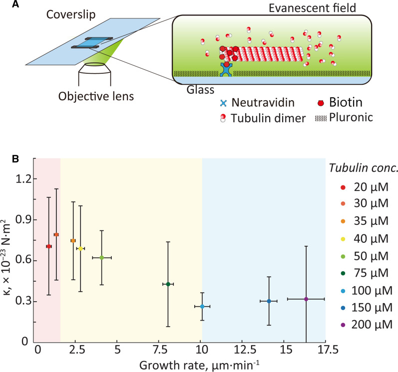
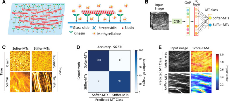
Results: Here, plausible explanations for these discrepancies are provided and a new method to evaluate the MT rigidity is developed. Moreover, a new relationship of the dynamic and mechanic of MTs is revealed that MT flexural rigidity decreases through three phases with the growth rate increases, which offers a method of designing MT flexural rigidity by regulating its growth rate. To test the validity of this method, the gliding performances of MTs with different flexural rigidities polymerized at different growth rates are examined. The growth rate-dependent flexural rigidity of MTs is experimentally found to influence the pattern formation in collective motion using gliding motility assay, which is further validated using machine learning.

Conclusion: Our study establishes a robust quantitative method for measurement and design of MT flexural rigidity to study its influences on MT gliding assays, collective motion, and other biological activities in vitro. The new relationship about the growth rate and rigidity of MTs updates current concepts on the dynamics and mechanics of MTs and provides comparable data for investigating the regulation mechanism of MT rigidity in vivo in the future.

Keywords: Collective motion; Flexural rigidity; Growth rate; Localization precision; Microtubule.


Conflict of interest statement

There are no conflicts to declare.

Figures

Fig. 1
Fig. 1
Flexural rigidity measurement of MTs. A Illustration of a partially biotinylated MT, that was immobilized onto the Au-stripe-patterned substrate with biotin–streptavidin bindings and the free segment fluctuated as a cantilever beam under Brownian motion. B Sequential images of a fluctuating MT (orange red) immobilized on the Au-stripe (light orange). Scale bar = 5 μm. Theoretical model of MT thermal fluctuation with localization precision of C 1 nm, D 10 nm, and E 100 nm. MT shapes in 500 frames are superimposed with different colors
Fig. 2
Fig. 2
Measurement process of κ for an MT of L = 10 µm and κset = 0.3 × 10−23 N·m2 with different localization precision. MT shapes (blue lines) and fitted curves (red lines and dots) with localization precision of A 1 nm and B 100 nm. Relationship between qn4 and L4/var(an) with localization precision of C 1 nm and D 100 nm. Blue dots: measurement results. Red lines: fitted straight lines
Fig. 3
Fig. 3
Error measurement of MT flexural rigidity under each condition of L, κset, and localization precision of A 1 nm, B 10 nm, and C 100 nm. Cross-hatched bars: L = 5 μm. Hatched bars: L = 10 μm. Shaded bars: L = 30 μm
Fig. 4
Fig. 4
Relationship between growth rate and flexural rigidity of MTs. A Growth rate measurement of MTs elongating under different tubulin concentrations as observed using TIRF microscopy. The partially biotinylated seed MTs were immobilized onto substrates via biotin–neutravidin bindings. Tubulin protein solution with a specific concentration was introduced into the flow cell and MT elongation process was observed directly. B MT flexural rigidity decreases with an increase in growth rate, which follows a three-state change and is illustrated with red, yellow, and blue background. The corresponding tubulin concentrations of the three stages are ≤ 30 μM, 30–100 μM, and > 100 μM, respectively. MTs incubated in tubulin concentrations from 20–200 μM are illustrated by the dots with different colors
Fig. 5
Fig. 5
Distinct patterns formed by MTs with different flexural rigidities. A MT gliding assays conducted within a reconstructed MT-kinesin system in the presence of methylcellulose. B A deep learning CNN model, including global average pooling (GAP) layer, final connected (FC) layer, and MT class output, is constructed to classify the two MTs: softer-MTs and stiffer-MTs. The two groups of MTs are polymerized at different tubulin concentration of 30 μM and 100 μM, which are named as softer-MTs (κ = 0.27 × 10−23 N m2) and stiffer-MTs (κ = 0.80 × 10−23 N m2), respectively. C From the identical isotropic state, softer-MTs and stiffer-MTs form distinctive patterns at the nematic phase. The addition time of ATP was set as 0 min. Scale bar = 50 μm. D Confusion matrix based on the trained CNN classifier. Here, 200 MT pattern images are categorized using the classifier. E The classification strategy of CNN classifier is visually explained using Score-CAM

References

    1. Mücke N, Kreplak L, Kirmse R, Wedig T, Herrmann H, Aebi U. Assessing the flexibility of intermediate filaments by atomic force microscopy. J Mol Biol. 2004;335:1241–1250. - PubMed
    1. Pallavicini C, Levi V, Wetzler DE, Angiolini JF, Benseñor L, Despósito MA. Lateral motion and bending of microtubules studied with a new single-filament tracking routine in living cells. Biophys J. 2014;106:2625–2635. - PMC - PubMed
    1. Fletcher DA, Mullins RD. Cell mechanics and the cytoskeleton. Nature. 2010;463:485–492. - PMC - PubMed
    1. Hawkins T, Mirigian M, Selcuk Yasar M, Ross JL. Mechanics of microtubules. J Biomech. 2010;43:23–30. - PubMed
    1. Sumino Y, Nagai KH, Shitaka Y, Tanaka D, Yoshikawa K, Chaté H. Large-scale vortex lattice emerging from collectively moving microtubules. Nature. 2012;483:448–452. - PubMed