

Claudin-7 deficiency promotes stemness properties in colorectal cancer through Sox9-mediated Wnt/β-catenin signalling
- PMID: 34281572
- PMCID: PMC8287764
- DOI: 10.1186/s12967-021-02983-3
Claudin-7 deficiency promotes stemness properties in colorectal cancer through Sox9-mediated Wnt/β-catenin signalling
Abstract
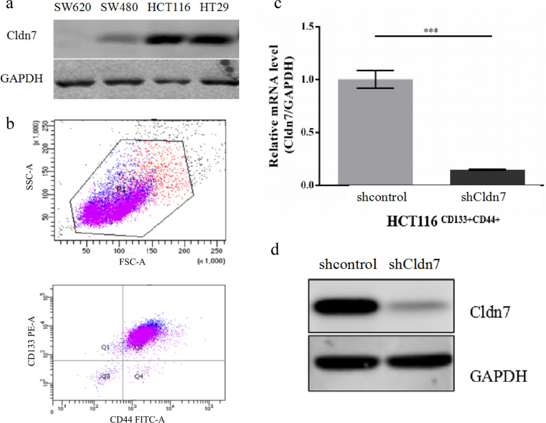
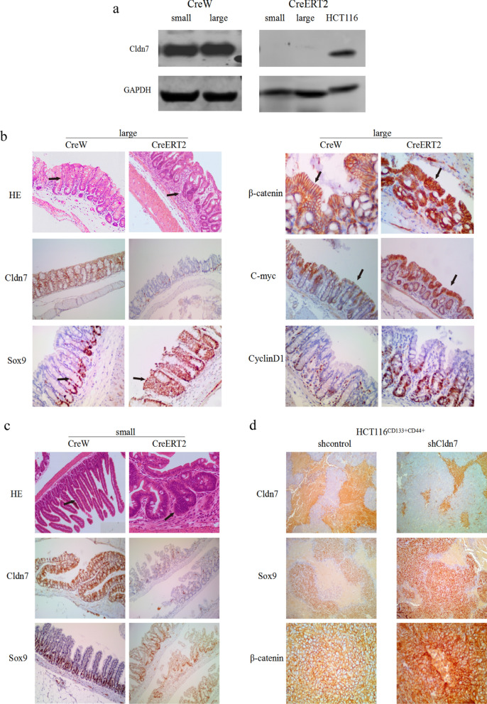
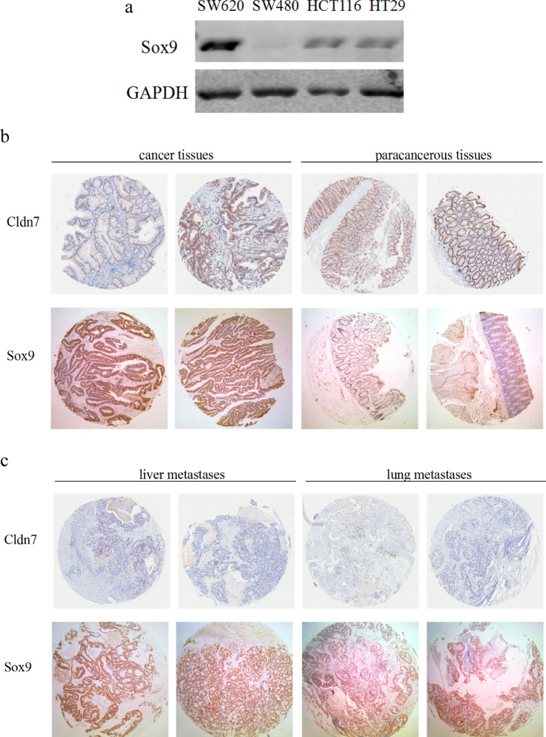
Background: Colorectal cancer (CRC) is a common malignant tumour of the digestive tract that is characterized by high patient morbidity and mortality rates. Claudin-7 (Cldn7), a tight junction protein, was recently reported to function as a candidate tumour suppressor gene in CRC. Our previous study demonstrated that the large intestine of C57/BL6 mice showed intestinal adenomas and abnormal Ki67 expression and distribution in the intestinal crypt when Cldn7 was knocked out. The aim of this study was to further investigate whether Cldn7 deficiency has non-tight junction functions, affects intestinal stemness properties, promotes CRC and to determine the specific mechanism.
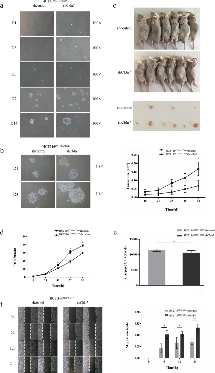
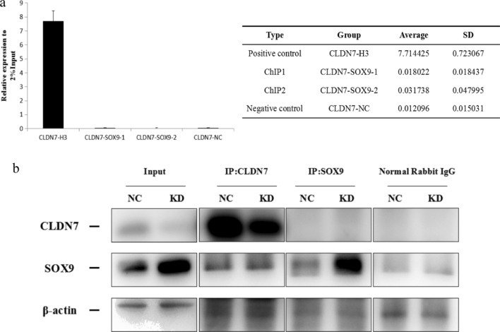
Methods: Cell proliferation assays, migration assays, apoptosis assays, tumour sphere formation assays in vitro, and subcutaneous xenograft models in vivo were used to determine the effects of Cldn7 knockdown on the biological characteristics of CRC stem cells. Western blotting, qPCR and immunofluorescence staining were performed to identify the epithelial-mesenchymal transition and the activation of Wnt/β-catenin pathway in CRC stem cells. Cldn7 inducible conditional gene knockout mice and immunohistochemical staining further verified this hypothesis in vivo. The mechanism and target of Cldn7 were determined by performing a chromatin immunoprecipitation (ChIP) assay and coimmunoprecipitation (CoIP) assay.
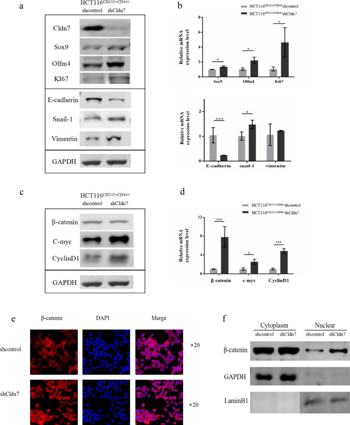
Results: Cldn7 knock down in CRC stem cells promoted cell proliferation, migration, and globular growth in serum-free medium and the ability to form xenograft tumours; cell apoptosis was inhibited, while the cellular epithelial-mesenchymal transition was also observed. These changes in cell characteristics were achieved by activating the Wnt/β-catenin pathway and promoting the expression of downstream target genes after β-catenin entry into the nucleus, as observed in CRC cell lines and Cldn7 gene knockout mouse experiments. Using ChIP and CoIP experiments, we initially found that Cldn7 and Sox9 interacted at the protein level to activate the Wnt/β-catenin pathway.
Conclusions: Based on our research, Cldn7 deficiency confers stemness properties in CRC through Sox9-mediated Wnt/β-catenin signalling. This result clarifies that Cldn7 plays an inhibitory role in CRC and reveals a possible molecular mechanism, which is conducive to further research on Cldn7 and cancer stem cells.
Keywords: Cancer stem cells; Claudin-7; Colorectal cancer; Epithelial–mesenchymal transition; Wnt/β-catenin.
© 2021. The Author(s).
Conflict of interest statement
The authors declare that they have no competing interests.
Figures
Similar articles
-
TM4SF1 promotes EMT and cancer stemness via the Wnt/β-catenin/SOX2 pathway in colorectal cancer.J Exp Clin Cancer Res. 2020 Nov 5;39(1):232. doi: 10.1186/s13046-020-01690-z. J Exp Clin Cancer Res. 2020. PMID: 33153498 Free PMC article.
-
RUNX1 promotes tumour metastasis by activating the Wnt/β-catenin signalling pathway and EMT in colorectal cancer.J Exp Clin Cancer Res. 2019 Aug 1;38(1):334. doi: 10.1186/s13046-019-1330-9. J Exp Clin Cancer Res. 2019. PMID: 31370857 Free PMC article.
-
Disheveled3 enhanced EMT and cancer stem-like cells properties via Wnt/β-catenin/c-Myc/SOX2 pathway in colorectal cancer.J Transl Med. 2023 May 5;21(1):302. doi: 10.1186/s12967-023-04120-8. J Transl Med. 2023. PMID: 37147666 Free PMC article.
-
Wnt/β-catenin signalling, epithelial-mesenchymal transition and crosslink signalling in colorectal cancer cells.Biomed Pharmacother. 2024 Jun;175:116685. doi: 10.1016/j.biopha.2024.116685. Epub 2024 May 5. Biomed Pharmacother. 2024. PMID: 38710151 Review.
-
Wnt signaling in colorectal cancer: pathogenic role and therapeutic target.Mol Cancer. 2022 Jul 14;21(1):144. doi: 10.1186/s12943-022-01616-7. Mol Cancer. 2022. PMID: 35836256 Free PMC article. Review.
Cited by
-
Single-cell analysis reveals landscape of endometrial cancer response to estrogen and identification of early diagnostic markers.PLoS One. 2024 Mar 22;19(3):e0301128. doi: 10.1371/journal.pone.0301128. eCollection 2024. PLoS One. 2024. PMID: 38517922 Free PMC article.
-
Colonic crypt stem cell functions are controlled by tight junction protein claudin-7 through Notch/Hippo signaling.Ann N Y Acad Sci. 2024 May;1535(1):92-108. doi: 10.1111/nyas.15137. Epub 2024 Apr 10. Ann N Y Acad Sci. 2024. PMID: 38598500 Free PMC article.
-
Advances of Wnt Signalling Pathway in Colorectal Cancer.Cells. 2023 Jan 30;12(3):447. doi: 10.3390/cells12030447. Cells. 2023. PMID: 36766788 Free PMC article. Review.
-
Claudin-7 is essential for the maintenance of colonic stem cell homoeostasis via the modulation of Wnt/Notch signalling.Cell Death Dis. 2024 Apr 23;15(4):284. doi: 10.1038/s41419-024-06658-x. Cell Death Dis. 2024. PMID: 38654000 Free PMC article.
-
Is High Expression of Claudin-7 in Advanced Colorectal Carcinoma Associated with a Poor Survival Rate? A Comparative Statistical and Artificial Intelligence Study.Cancers (Basel). 2022 Jun 13;14(12):2915. doi: 10.3390/cancers14122915. Cancers (Basel). 2022. PMID: 35740581 Free PMC article.
References
-
- Lu Z, Liu Y, Xu J, Yin HP, Yuan HY, Gu JJ, et al. Immunohistochemical quantification of expression of a tight junction protein, claudin-7, in human lung cancer samples using digital image analysis method. Comput Methods Programs Biomed. 2018;155:179–187. doi: 10.1016/j.cmpb.2017.12.014. - DOI - PubMed
-
- Bernardi MA, Logullo AF, Pasini FS, Nonogaki S, Blumke C, Soares FA, et al. Prognostic significance of CD24 and claudin-7 immunoexpression in ductal invasive breast cancer. Oncol Rep. 2012;27:28–38. - PubMed
Publication types
MeSH terms
Substances
LinkOut - more resources
Full Text Sources
Medical
Research Materials
