. 2021 Oct 15;90(8):563-574.
doi: 10.1016/j.biopsych.2021.05.022. Epub 2021 Jun 1.

Hypoactive Thalamic Crh+ Cells in a Female Mouse Model of Alcohol Drinking After Social Trauma

Affiliations

Hypoactive Thalamic Crh+ Cells in a Female Mouse Model of Alcohol Drinking After Social Trauma

Emily L Newman et al. Biol Psychiatry. .

Abstract

Background: Comorbid stress-induced mood and alcohol use disorders are increasingly prevalent among female patients. Stress exposure can disrupt salience processing and goal-directed decision making, contributing to persistent maladaptive behavioral patterns; these and other stress-sensitive cognitive and behavioral processes rely on dynamic and coordinated signaling by midline and intralaminar thalamic nuclei. Considering the role of social trauma in the trajectory of these debilitating psychopathologies, identifying vulnerable thalamic cells may provide guidance for targeting persistent stress-induced symptoms.

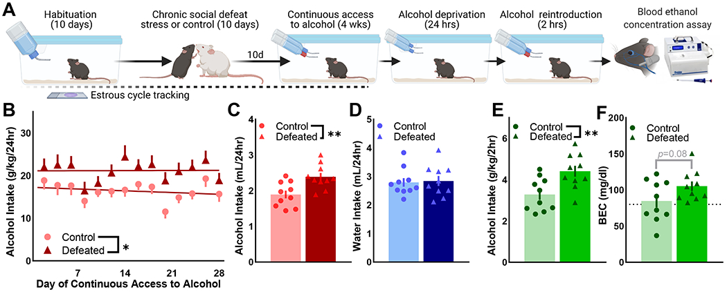
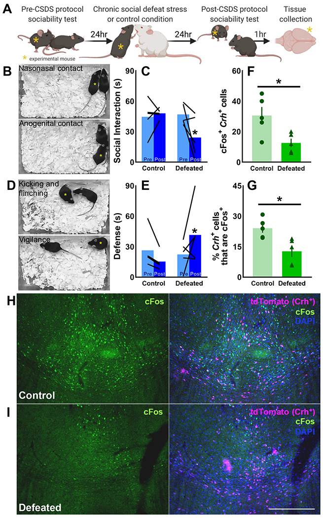
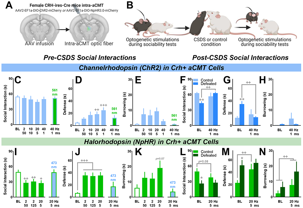
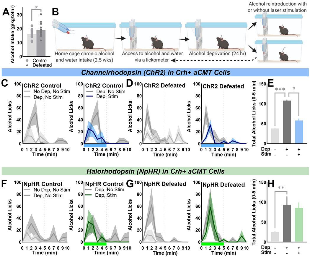
Methods: A novel behavioral protocol traced the progression from social trauma to the development of social defensiveness and chronically escalated alcohol consumption in female mice. Recent cell activation-measured as cFos-was quantified in thalamic cells after safe social interactions, revealing stress-sensitive corticotropin-releasing hormone-expressing (Crh+) anterior central medial thalamic (aCMT) cells. These cells were optogenetically stimulated during stress-induced social defensiveness and abstinence-escalated binge drinking.

Results: Crh+ aCMT neurons exhibited substantial activation after social interactions in stress-naïve but not in stressed female mice. Photoactivating Crh+ aCMT cells dampened stress-induced social deficits, whereas inhibiting these cells increased social defensiveness in stress-naïve mice. Optogenetically activating Crh+ aCMT cells diminished abstinence-escalated binge alcohol drinking in female mice, regardless of stress history.

Conclusions: This work uncovers a role for Crh+ aCMT neurons in maladaptive stress-induced social interactions and in binge drinking after forced abstinence in female mice. This molecularly defined thalamic cell population may serve as a critical stress-sensitive hub for social deficits caused by exposure to social trauma and for patterns of excessive alcohol drinking in female populations.

Keywords: Alcohol; Corticotropin-releasing hormone; Defensive behavior; Female chronic social defeat stress; Social interaction; Thalamus.


Conflict of interest statement

Disclosures: The authors report no biomedical financial interests or potential conflicts of interest.

Figures

Figure 1.
Figure 1.. Social stress and forced abstinence increase alcohol drinking in female mice.
(A) Wild-type C57BL/6J female mice were exposure to 10-day chronic social defeat stress (CSDS) or the non-defeated control condition (n=10/condition). Ten days after the final day of CSDS, mice received four weeks of continuous access to 20% ethyl alcohol (EtOH (w/v)) and water in the home cage. Alcohol was removed for 24-hours and reintroduced for 2-hours; submandibular blood was collected to determine blood EtOH concentration (BEC). Estrous cycle phase was tracked throughout (Fig. S1). (B) Defeated female mice consistently consumed more alcohol (g/kg/24hr) than control mice in the four weeks of alcohol access [F(1,18)=8.29, p=0.01]; data depicted as two-day group Means ± SEM. (C) Average daily alcohol intake was greater in defeated mice compared to controls [t(18)=3.22, p=0.0048] whereas (D) water intake was similar across conditions. (E) Defeated mice consumed more alcohol during two-hour access after forced abstinence [t(18)=3.065, p=0.0067], (F) producing a trend of increased BEC in defeated vs. control mice (p=0.08). Average 2-hour BECs were > 80 mg/dL (dotted line), indicative of a binge pattern of alcohol intake in control and defeated mice. (C, D) Data points depict four-week averages for individual mice; bars represent four-week group Mean ± SEM. (E, F) Data points are individual mice; bars represent the group Mean ± SEM. * p <0.05, ** p<0.01 control vs. defeated
Figure 2.
Figure 2.. Chronic social defeat stress blunts social cFos activation in Crh+ aCMT cells.
(A) Female CRH-ires-Cre/Ai9 mice were tested for baseline sociability toward a safe social partner, then exposed to either 10-day chronic social defeat stress (CSDS; n=5 Defeated) or the 10-day control condition (n=5 Control. Sociability was retested the day after 10-day CSDS/control protocol; tissue was collected one hour later to examine colocalization of cFos with the endogenous Crh reporter protein, tdTomato, in anterior central medial thalamic (aCMT) cells. (B, C) CSDS reduced sociability, measured as cumulative nasonasal and anogenital contact time [F(1,8)=5.37, p=0.049] and (D, E) increased defensiveness, measured as total time spent kicking, flinching, and maintaining a vigilant-like pose [F(1,8)=5.56, p=0.046]. (B, D) Yellow asterisks indicate CRH-ires-Cre/Ai9 mice as they engage in representative social and defensive behaviors. (F) Colocalization of cFos and tdTomato in the aCMT was suppressed by CSDS [t(8)=2.87, p=0.021]. (G) A greater percentage of Crh+ aCMT cells were activated by safe social interactions in (H) control mice compared to (I) defeated individuals [t(8)=3.19, p=0.013]; (H, I) representative images taken at 10x magnification of (left panels) cFos (green) and (right panels) cFos colocalization with tdTomato (magenta) and DAPI counterstaining (blue); 200 μm scale bar. Data shown as individual values and as the group Mean ± SEM; *p<0.05 control vs. defeated
Figure 3.
Figure 3.. Optogenetic activation of Crh+ aCMT neurons reverses chronic social defeat stress-induced social deficits whereas optogenetic inhibition produces a defeat-like phenotype in stress-naïve female mice.
(A) Female CRH-ires-Cre mice received intra-aCMT AAV to express Cre-dependent channelrhodopsin (ChR2) or halorhodopsin (NpHR) in Crh+ neurons and were implanted with optic fibers targeting the aCMT. (B) Laser pulses were delivered during home cage safe social interactions to activate (ChR2) or inhibit (NpHR) Crh+ aCMT cells before and after 10-day chronic social defeat stress (CSDS) or the 10-day control procedure (ChR2: n=8 Control, n=10 Defeated; NpHR: n=8 Control, n=8 Defeated). (C) Before 10-day CSDS, neither 473 nm laser pulses nor control 561 nm laser pulses affected social interaction times in ChR2-expressing mice as compared with no-laser baseline (BL) tests. (D) Optogenetic activation of Crh+ aCMT cells produced a frequency-dependent increase in social defensiveness [F(4,68)=6.25, p<0.001] without affecting (E) burrowing. (F-H; BL) Compared with control mice, CSDS suppressed sociability [t(16)=4.35, p<0.001] and increased defensiveness [t(16)=3.70, p=0.002] without affecting burrowing. In defeated mice, Crh+ aCMT cell activation (F) recovered sociability [F(1,16)=11.63, p=0.004], and (G) diminished defensiveness [F(1,16)=9.00, p=0.009] without influencing (H) burrowing. (I) Compared to BL tests, inhibiting Crh+ aCMT cells with 561 nm light pulses reduced the duration of social interactions [F(3,45)=7.61, p<0.001], (J) increased defensive behaviors [F(3,45)=12.24, p<0.001], and (K) increased time spent burrowing [F(3,45)=4.07, p=0.012] whereas 473 nm pulses had no effect. (L-N) Post-CSDS, defeated mice interacted less with a social partner compared to control mice [t(14)=2.61, p=0.021] and were more defensive [t(14)=2.94, p=0.011]. Optogenetic inhibition of Crh+ aCMT cells produced a defeat-like phenotype in control mice by (F) increasing defensiveness [F(1,14)=5.35, p=0.036] while also (G) increasing time spent burrowing [F(1,14)=7.25, p=0.018]. (C-N) Data shown as the Mean ± SEM; Defeat vs. control: *p<0.05, **p<0.01; no stimulation (BL) vs. stimulation: +p<0.05, ++p<0.01, +++p<0.001. (A) Coronal brain images adapted from the Allen Institute Mouse Brain Atlas
Figure 4.
Figure 4.. Optogenetic activation of Crh+ aCMT neurons diminishes abstinence-induced binge alcohol drinking in female mice.
(A) Chronically socially defeated female CRH-ires-Cre mice drank more alcohol (g/kg/24hr) than stress-naïve controls [one-tailed: t(33)=1.99, p=0.027] when given (B) continuous access to water (blue bottle) and 20% ethyl alcohol ((w/v); red bottle) in their home cage. Cages were fitted with contact lickometer panels to count alcohol and water bottle licks during unrestricted access (No Dep). Mice were alcohol-deprived (Dep) for 24 hours prior to alcohol reintroduction and optogenetic activation (ChR2) or inhibition (NpHR) of Crh+ aCMT neurons (Dep, Stim) or during sessions without laser stimulations (Dep, No Stim). (C, D, F, G) Average alcohol licks/minute over the first 10 minutes of each drinking session; data shown as the group Mean ± SEM (line ± shaded areas) with light grey lines indicating No Dep, No Stim sessions, dark grey lines indicating Dep, No Stim sessions, blue or green lines denoting Dep, Stim of ChR2 or NpHR sessions, respectively. Blue or green bars beneath x-axes indicate 5-minute stimulation of ChR2 or NpHR, respectively. Vertical dotted lines indicate laser stimulation offset or the matching time point during No Stim sessions. (C, D; left panels) Control (n=8) and defeated (n=10) ChR2-expressing female mice demonstrated deprivation-escalated alcohol drinking during the first five minutes of drinking during Dep vs. No Dep sessions [F(1,16)=17.37, p<0.001]; (C, D; right panels) optogenetic activation of Crh+ aCMT neurons blunted deprivation-induced binge drinking in both control and defeated mice [F(1,16) = 5.72, p=0.029]. (E) Summarized effects of deprivation and Crh+ aCMT cell activation on the first five minutes of alcohol intake; data shown as Mean ± SEM collapsed over control and defeat conditions. (F, G; left panels) Control (n=9) and defeated (n=8) NpHR-expressing mice exhibited deprivation-escalated alcohol drinking during the first five minutes of Dep vs. No Dep sessions [F(1,15) =11.33, p=0.004]. (F, G, right panels) Optogenetic inhibition of Crh+ aCMT neurons had no effect on deprivation-escalated intake. (H) Summary of effects of alcohol deprivation and optogenetic inhibition on alcohol licking during the first five minutes of each session type in NpHR-expressing mice; data depicted as Mean ± SEM collapsed over control and defeat groups. **p<0.01, ***p<0.001 deprivation effect; # p<0.05 stimulation effect.

Comment in

Similar articles

Cited by

References

    1. Kessler RC (2003): Epidemiology of women and depression. J Affect Disord. 74:5–13. - PubMed
    1. Kessler RC (1997): The effects of stressful life events on depression. Annu Rev Psychol. 48:191–214. - PubMed
    1. Kessler RC, Sonnega A, Bromet E, Hughes M, Nelson CB (1995): Posttraumatic stress disorder in the National Comorbidity Survey. Arch Gen Psychiatry. 52:1048–1060. - PubMed
    1. Kessler RC, McGonagle KA, Swartz M, Blazer DG, Nelson CB (1993): Sex and depression in the National Comorbidity Survey. I: Lifetime prevalence, chronicity and recurrence. J Affect Disord. 29:85–96. - PubMed
    1. Breslau N, Davis GC, Andreski P, Peterson EL, Schultz LR (1997): Sex differences in posttraumatic stress disorder. Arch Gen Psychiatry. 54:1044–1048. - PubMed

Publication types

Substances