Review
. 2022 Feb;47(2):124-135.
doi: 10.1016/j.tibs.2021.06.008. Epub 2021 Jul 16.

Drug discovery in the era of cryo-electron microscopy

Affiliations
Review

Drug discovery in the era of cryo-electron microscopy

Michael J Robertson et al. Trends Biochem Sci. 2022 Feb.

Abstract

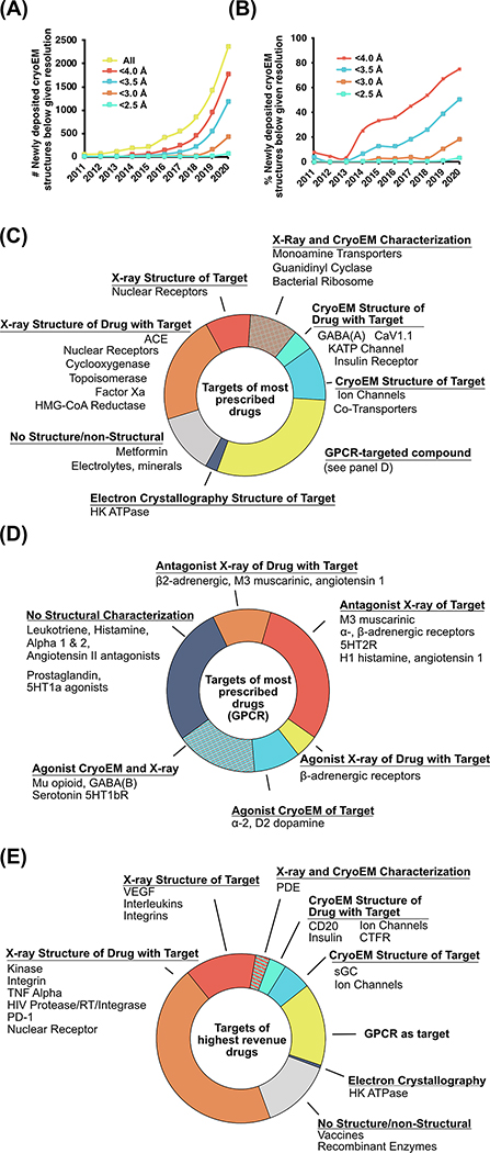
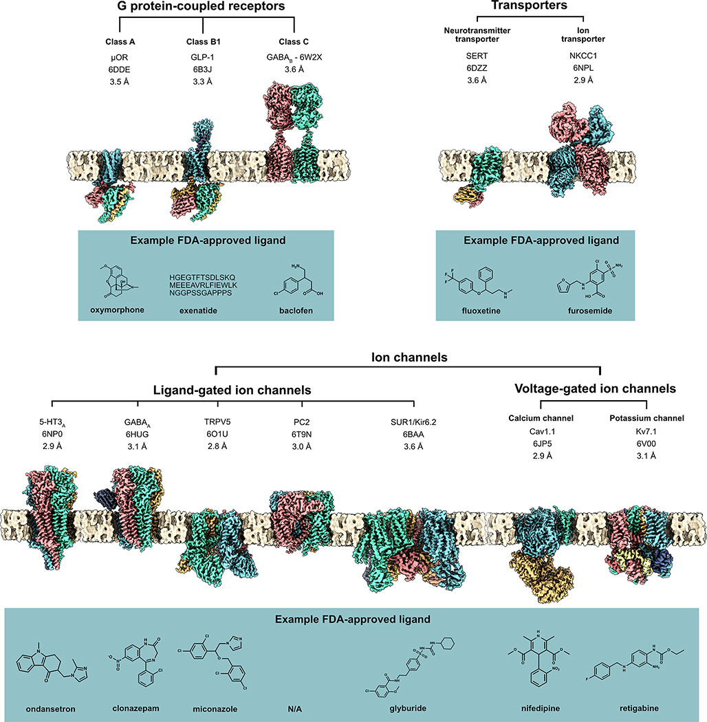
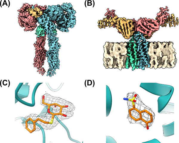
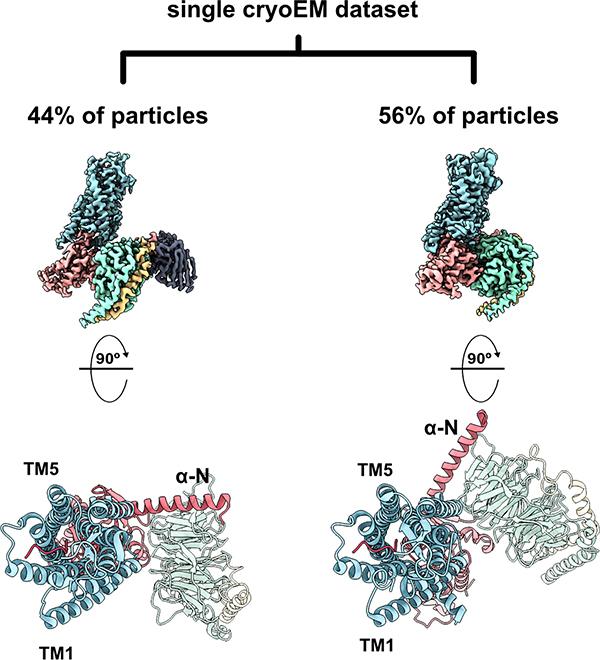
Structure-based drug discovery (SBDD) is an indispensable approach for the design and optimization of new therapeutic agents. Here, we highlight the rapid progress that has turned cryo-electron microscopy (cryoEM) into an exceptional SBDD tool, and the wealth of new structural information it is providing for high-value pharmacological targets. We review key advantages of a technique that directly images vitrified biomolecules without the need for crystallization; both in terms of a broader array of systems that can be studied and the different forms of information it can provide, including heterogeneity and dynamics. We discuss near- and far-future developments, working in concert towards achieving the resolution and throughput necessary for cryoEM to make a widespread impact on the SBDD pipeline.

Keywords: biologics; cryo-electron microscopy; pharmacology; single-particle analysis; small molecule; structure-based drug discovery.


Conflict of interest statement

Declaration of interests The authors declare that they have no conflicts of interest with the content of this article.

Figures

Figure 1:
Figure 1:. The improvement in resolution of cryoEM and its contribution to the structural characterization of protein drug targets.
A) The absolute number of newly deposited cryoEM structures in the PDB below given resolutions and B) percentage of newly deposited cryoEM structures in the PDB below given resolutions [61]. C) Chart of the targets of the 200 most-prescribed drugs of 2018 broken down by the target’s structural characterization, D) chart of the targets of the 44 most-prescribed drugs targeting GPCRs broken down by structural characterization, and E) chart of the targets of the 200 highest-sales drugs (as a proxy for newer drugs) of 2018 broken down by the target’s structural characterization. Data manually curated in 2020 by identification of the protein target, if applicable, of the 200 most prescribed and 200 highest sales drugs as curated by the Njardarson lab for 2019 [14], followed by the identification of related structures in the PDB.
Figure 2, Key Figure:
Figure 2, Key Figure:. High-resolution cryoEM maps of a wide variety of membrane protein drug targets.
This includes G protein-coupled receptors (GPCRs) and transporters (top row) and ion channels (bottom row), with an example FDA-approved ligand for each receptor (blue boxes). PDB IDs and resolution provided below protein name. Membrane generated with CHARMM-GUI [62] and figure rendered in ChimeraX [63]. GABAA (PDB ID 6HUG) and TRPV5 (PDB ID 6O1U) were determined in lipid nanodiscs; all others displayed were obtained in detergent micelles.
Figure 3:
Figure 3:. Demonstration of the utility of cryoEM for both small-molecule and biologics discovery.
CryoEM maps of (A) the insulin receptor bound to insulin (PDB ID 6PXV) and (B) CD20 in complex with rituximab Fabs (PDB ID 6VJA). (C) Accurate modeling of PETG into a cryoEM map of β-galactosidase using GemSpot (PDB ID 6CVM). (D) Fragment-based discovery for PKM2 with cryoEM density allowing for correct identification and placement of discovery fragment (PDB ID 6TTF).
Figure 4:
Figure 4:. From a single cryoEM dataset, 3-dimensional classification of projections revealed two distinct conformers representing two distinct GPCR-G protein interaction states representing two thermodynamically comparable conformers.
In the canonical state (left, PDB ID 6OS9), the receptor engages the G protein in a prototypical fashion in which the nucleotide binding pocket is primed for GTP binding. In the non-canonical state (right, PDB ID 6OSA), the G protein heterotrimer is rotated by 45° compared to the canonical state, representing an intermediate ligand-bound receptor state along the G protein coupling pathway. TM: transmembrane helix; α-N: N-terminal alpha helix of G protein.

References

    1. Byers LD and Wolfenden R (1972) A potent reversible inhibitor of carboxypeptidase A. J. Biol. Chem. 247, 606–608 - PubMed
    1. Patlak M (2004) From viper’s venom to drug design: treating hypertension. FASEB J. 18, 421. - PubMed
    1. Baldwin JJ et al. (1989) Thienothiopyran-2-sulfonamides: novel topically active carbonic anhydrase inhibitors for the treatment of glaucoma. J. Med. Chem. 32, 2510–2513 - PubMed
    1. Eder J et al. (2014) The discovery of first-in-class drugs: origins and evolution. Nat. Rev. Drug Discov. 13, 577–587 - PubMed
    1. Ostrem JM et al. (2013) K-Ras(G12C) inhibitors allosterically control GTP affinity and effector interactions. Nature 503, 548–551 - PMC - PubMed

Publication types

MeSH terms

LinkOut - more resources