

A Unique Technique for Precise Targeting in Treatment of Rare Bifocal Intraosseous Ganglion Cysts of the Talus: A Case Report and Review of the Literature
- PMID: 34282110
- PMCID: PMC8311387
- DOI: 10.12659/AJCR.932261
A Unique Technique for Precise Targeting in Treatment of Rare Bifocal Intraosseous Ganglion Cysts of the Talus: A Case Report and Review of the Literature
Abstract
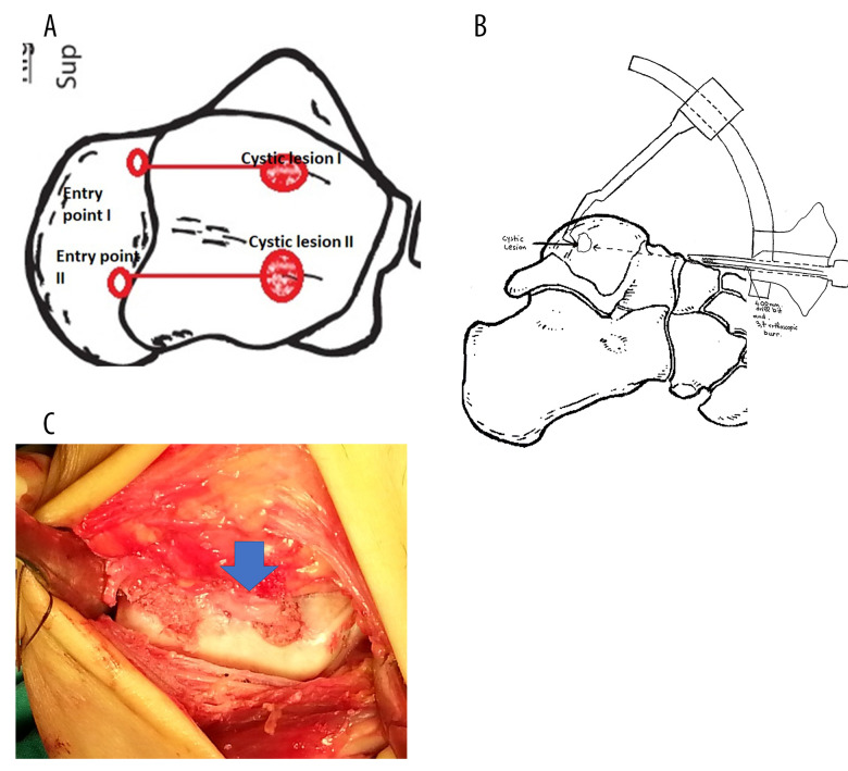
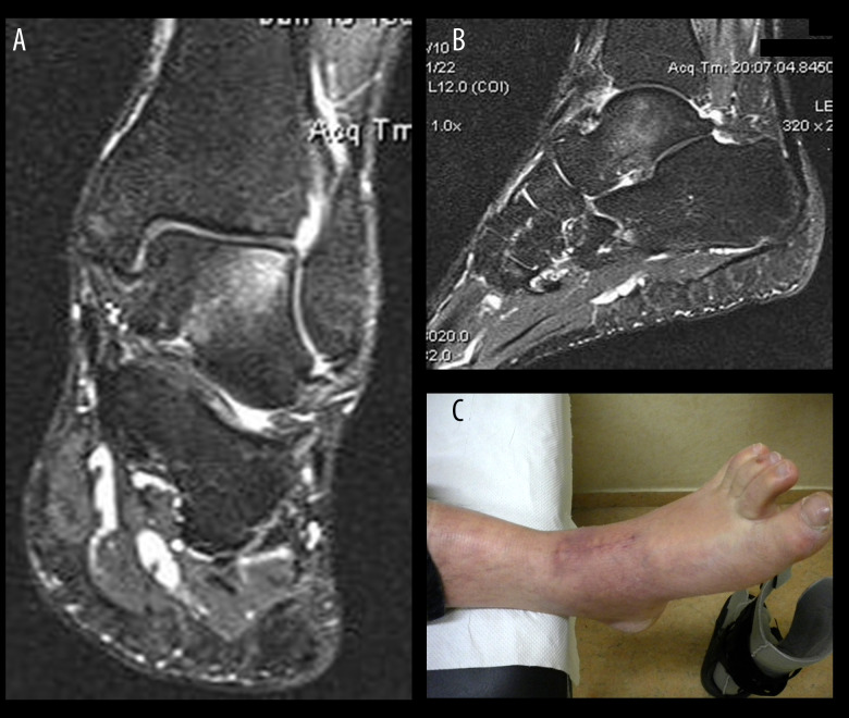
BACKGROUND This article presents a rare case of 2 separate intraosseous ganglion cysts of the talus in a 51-year-old man, treated with a unique technique of precise lesion targeting to avoid extensive bone loss and minimize articular chondral injury of the talus. CASE REPORT Two separate intraosseous ganglion cysts of the talus were diagnosed in a 51-year-old man with chronic ankle pain. A single straight-line incision with an entry point through the talonavicular joint was created to spare the precarious blood supply of the talus network. The 2 distinct subchondral lesions were approached under fluoroscopic control for curettage and autologous bone grafting using the anterior cruciate ligament tibial guide in a pair-of-compasses fashion. In almost 5 years of follow-up the patient has been asymptomatic. Magnetic resonance imaging has revealed no signs of degenerative changes in the ankle or the talonavicular joint, and the intraosseous edema has almost disappeared. CONCLUSIONS To the best of our knowledge, this case is the first report of 2 distinct intraosseous ganglion cysts of the talus. We recommend the precise targeting technique used in our case for treating intraosseous talar lesions with intact articular cartilage.
Conflict of interest statement
None.
Figures


References
-
- Lorkowski J, Mrzygłód MW, Grzegorowska O, Kotela I. An in silico analysis of ankle joint loads in secondary ankle osteoarthritis. Case study. Ortop Traumatol Rehabil. 2015;17(3):305–15. - PubMed
-
- Nishimura T, Tsujii M, Kusuzaki K, et al. Intra-osseous ganglion of the proximal humerus: A case report. J Orthop Surg (Hong Kong) 2007;15(1):102–5. - PubMed
-
- Buldu H, Kantarci U, Cepel S. [Intraosseous ganglions at the same localization in twin sisters.] Acta Orthop Traumatol Turc. 2009;43(4):379–80. [in Turkish] - PubMed
Publication types
MeSH terms
LinkOut - more resources
Full Text Sources
