. 2021 Aug;6(8):1031-1042.
doi: 10.1038/s41564-021-00937-5. Epub 2021 Jul 19.

MX2-mediated innate immunity against HIV-1 is regulated by serine phosphorylation

Affiliations

MX2-mediated innate immunity against HIV-1 is regulated by serine phosphorylation

Gilberto Betancor et al. Nat Microbiol. 2021 Aug.

Erratum in

Abstract

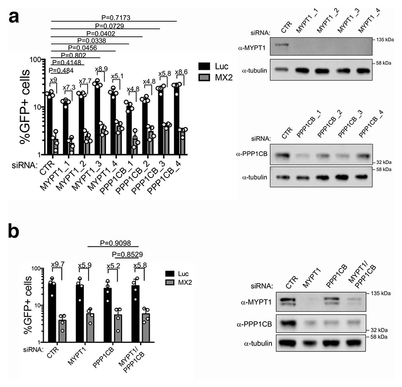
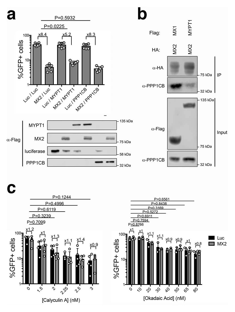
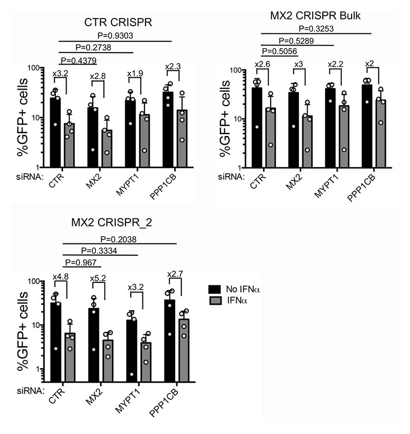
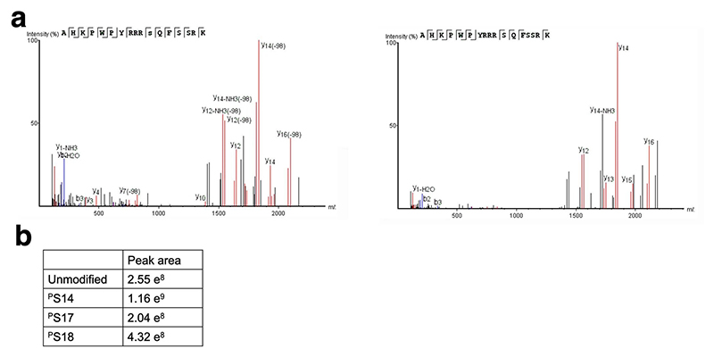
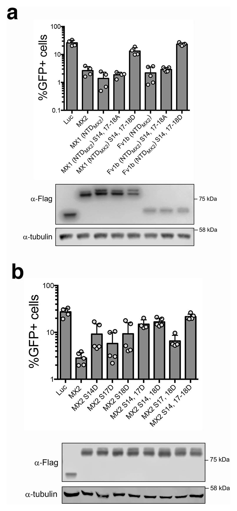
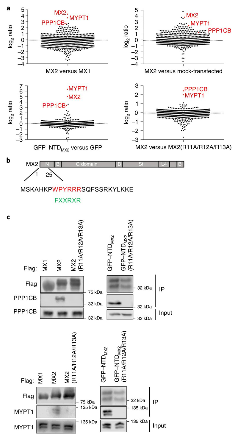
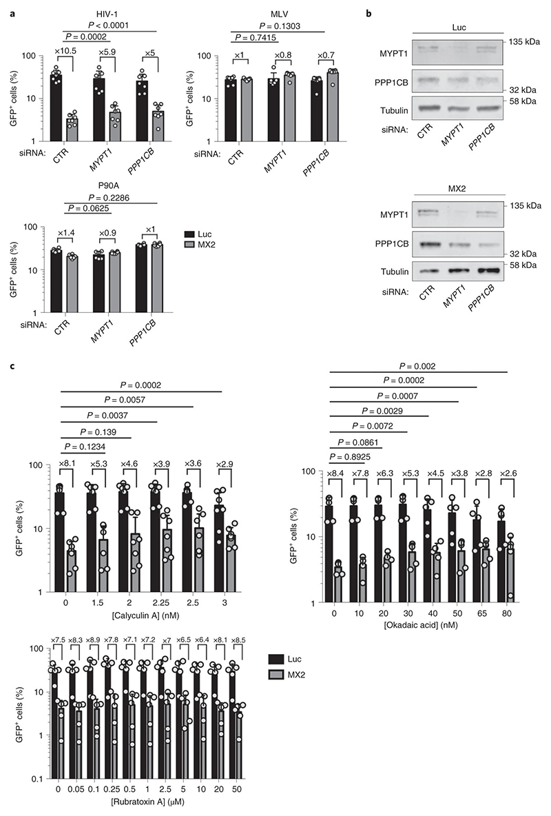
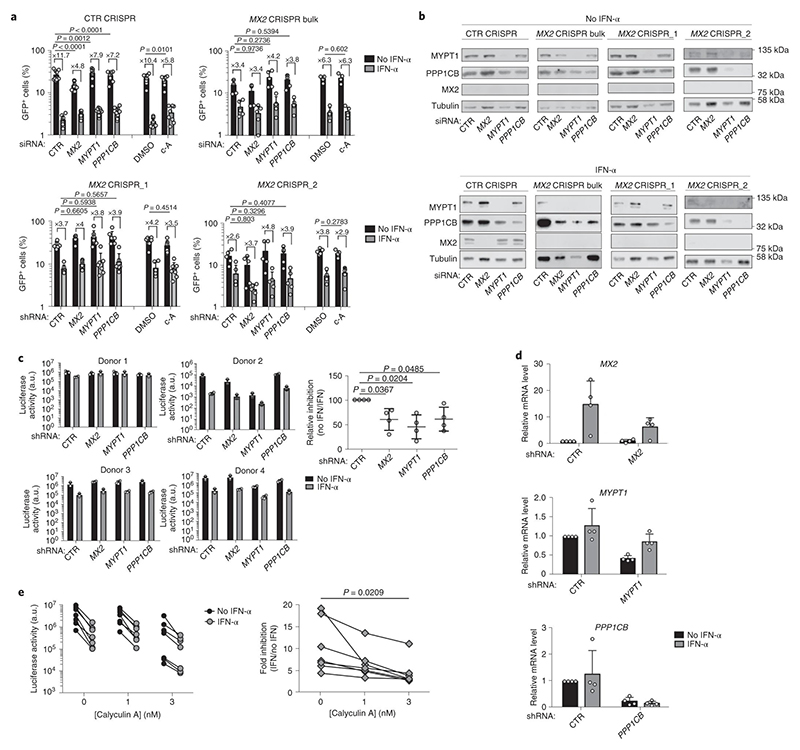
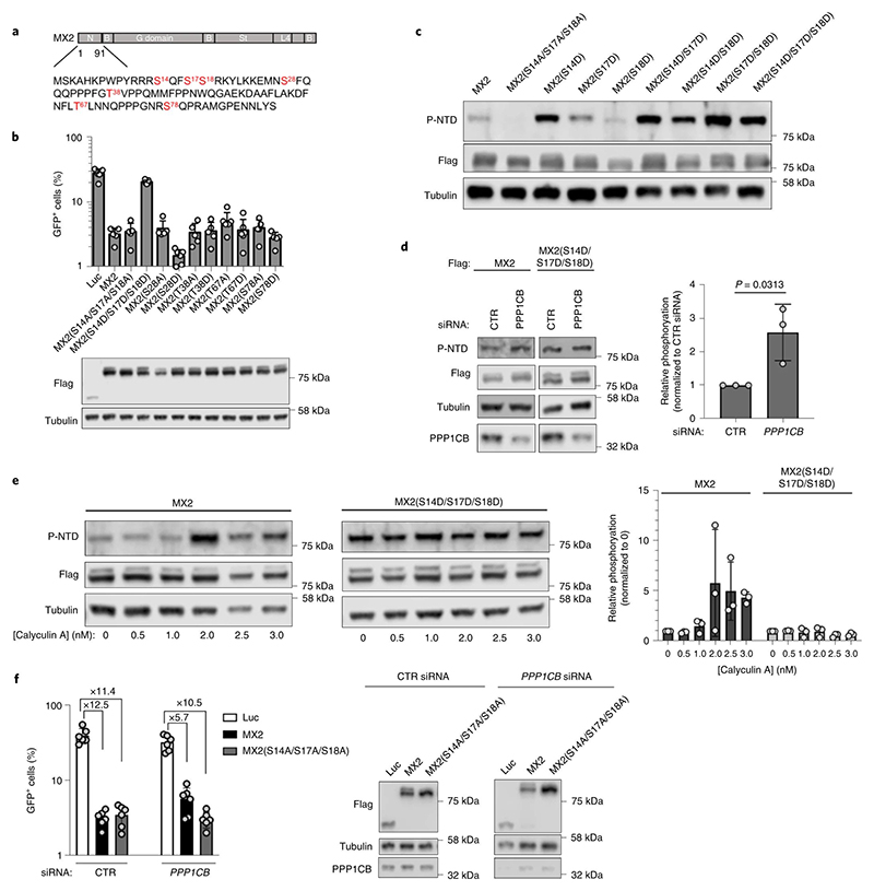
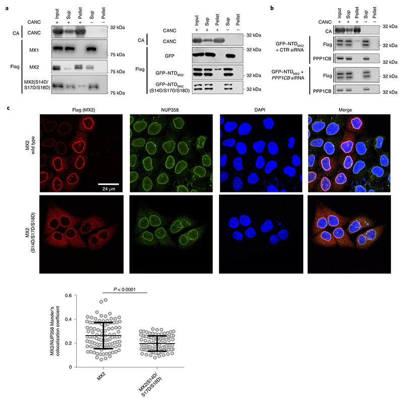
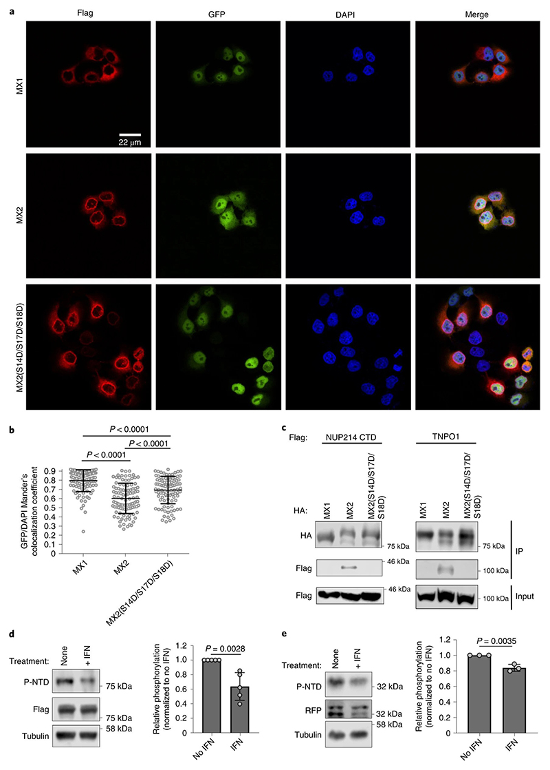
The antiviral cytokine interferon activates expression of interferon-stimulated genes to establish an antiviral state. Myxovirus resistance 2 (MX2, also known as MxB) is an interferon-stimulated gene that inhibits the nuclear import of HIV-1 and interacts with the viral capsid and cellular nuclear transport machinery. Here, we identified the myosin light chain phosphatase (MLCP) subunits myosin phosphatase target subunit 1 (MYPT1) and protein phosphatase 1 catalytic subunit-β (PPP1CB) as positively-acting regulators of MX2, interacting with its amino-terminal domain. We demonstrated that serine phosphorylation of the N-terminal domain at positions 14, 17 and 18 suppresses MX2 antiviral function, prevents interactions with the HIV-1 capsid and nuclear transport factors, and is reversed by MLCP. Notably, serine phosphorylation of the N-terminal domain also impedes MX2-mediated inhibition of nuclear import of cellular karyophilic cargo. We also found that interferon treatment reduces levels of phosphorylation at these serine residues and outline a homeostatic regulatory mechanism in which repression of MX2 by phosphorylation, together with MLCP-mediated dephosphorylation, balances the deleterious effects of MX2 on normal cell function with innate immunity against HIV-1.


Conflict of interest statement

Conflict of interest

The authors declare that they have no conflict of interest.

Figures

Extended Data Fig. 1
Extended Data Fig. 2
Extended Data Fig. 3
Extended Data Fig. 4
Extended Data Fig. 5
Extended Data Fig. 6
Fig. 1
Fig. 1. The N-terminal domain of MX2 interacts with MYPT1 and PPP1CB
a, Stable isotope labelling by amino acids (SILAC) screen for MX2 binding proteins. Streptavidin-tagged MX2, MX1, MX2 R11-13A, GFP or GFP (NTDMX2) were expressed in 293T cells labelled with “light”, “medium” or “heavy” amino acids, immune-affinity purified and analyzed by tandem mass spectrometry. Representative experiments comparing MX2 (medium) versus MX1 (light) (n= 3 biological replicates), MX2 (medium) versus mock (light) transfected cells (n= 1), GFP (NTDMX2) (heavy) versus GFP (light) (n= 2 biological replicates) and MX2 (light) versus MX2 R11-13A (medium) (n= 3 biological replicates) are shown. Values on the y-axis are in Log-2 scale. b, MX2 domain organization showing the sequence of the N-terminal 25 amino acids, highlighting the MLCP binding motif (in red) and a consensus PP1 binding sequence (in green). c, Interaction of endogenous PPP1CB (upper panel) or MYPT1 (lower panel) with the MX2 N-terminal domain. Transfected 293T cells expressing Flag-tagged MX1, MX2, MX2 R11-13A, GFP (NTDMX2) or GFP (NTDMX2) R11-13A were lysed, proteins immunoprecipitated (IP) with an anti-Flag antibody and analyzed by immunoblot using anti-PPP1CB, anti-MYPT1 or anti-Flag antibodies. All experiments were done at least 4 times.
Fig. 2
Fig. 2. MX2 requires functional MLCP for antiviral activity
a, U87-MG CD4+ CXCR4+ cells were transduced with pEasiLV vectors expressing MX2 or the negative control protein Luc and protein expression induced by doxycycline (0.5 μg/ml) for 48 h. MYPT1 or PPP1CB were then depleted by double siRNA transfection. Cells were challenged with wild type or P90A versions of HIV-1/GFP or MLV/GFP and the percentage of infected cells from the pEasiLV transduced population (typically >85%), enumerated at 48 h by flow cytometry (n = 7 biological replicates for HIV-1; n = 6 biological replicates for P90A and MLV, mean ± standard deviation (SD) *p value <0.05; two-tailed unpaired t-test). b, Immunoblot analysis of MYPT1 and PPP1CB depletion in U87-MG CD4+ CXCR4+ cells from panel A. Tubulin is included as a loading control. c, U87-MG CD4+ CXCR4+ cells expressing MX2 or Luc were treated with increasing concentrations of calyculin A, okadaic acid or rubratoxin A (or DMSO as a control) 6 h before challenge with HIV-1/GFP. Inhibitors were removed 24 h after addition and the percentage of infected cells enumerated at 48 h by flow cytometry (n = 7 biological replicates for calyculin A; n = 5 biological replicates for okadaic acid and rubratoxin A, mean ± SD, *p value <0.05, (ns) non-significant; two-tailed unpaired t-test).
Fig. 3
Fig. 3. Interferon-induced MX2-mediated inhibition of HIV-1 requires MLCP
a, U87-MG CD4+ CXCR4+ cells transduced with a control guide RNA, as well as a bulk population and two clonal cell lines where MX2 had been disrupted by CRISPR-Cas9 genome editing, were either transfected twice with a control siRNA (CTR), or siRNAs targeting MX2, MYPT1 or PPP1CB, and treated or not with 500 U/ml IFNα for 24 h. Alternatively, cells were treated with 3 nM calyculin A (or DMSO) 6 h before infection and medium exchange 18 h later. Cells were challenged with HIV-1/GFP and infection enumerated at 48 h by flow cytometry (n = 10 biological replicates for CTR CRISPR cells siRNA treatment and n = 8 biological replicates for calyculin A treatment; n = 5 biological replicates for MX2 CRISPR Bulk cells; n = 7 biological replicates for MX2 CRISPR_1 cells; n = 5 biological replicates for MX2 CRISPR_2 cells, mean ± SD, *p value <0.05; two-tailed unpaired t-test). b, Immunoblot analysis of MX2, MYPT1, PPP1CB and tubulin (loading control) in CTR CRISPR, MX2 CRISPR Bulk, MX2 CRISPR_1 and MX2 CRISPR_2 cells (from panel A). c, Primary CD4+ T cells were isolated from 4 donors, transduced with shRNAs targeting MX2, MYPT1, PPP1CB or a scrambled sequence shRNA (CTR) and treated or not with 2,000 U/ml IFNα. At 24 h, cells were challenged with NL4.3/Nef-IRES-Renilla and luciferase activity determined 48 h later. Raw infectivity data are shown on the left (mean values from 3 technical replicates), and the fold inhibition of infection (no IFN/IFN) normalized to the CTR shRNA (arbitrary value of 100), on the right (*p-value <0.05; two-tailed paired t-test). d, MX2, MYPT1 or PPP1CB depletion in primary CD4+ T cells after shRNA transduction was quantitated by qPCR, normalizing to GAPDH. Data shown represent the donors used in panel C (n = 4, mean ± SD). e, Primary CD4+ T cells treated or not with 2,000 U/ml of IFNα were incubated in the presence of DMSO or two concentrations of calyculin A (1 nm or 3 nM). After 6 h, cells were challenged with NL4.3/Nef-IRES-Renilla and luciferase activity determined after 48 h. Raw infectivity data from all 7 donors are shown on the left and the fold inhibition of infection (no IFN/IFN), on the right (*p-value <0.05; two-tailed paired t-test).
Fig. 4
Fig. 4. MX2 antiviral activity is antagonized by phosphorylation
a, MX2 domain organization showing the sequence of the N-terminal domain, and indicating phosphorylated residues (in red). b, U87-MG CD4+ CXCR4+ cells were transduced with pEasiLV expressing Flag-tagged wild type or mutant MX2, or the Luc control. After induction with 0.5 μg/ml of doxycycline for 48 h, cells were challenged with HIV-1/GFP and infectivity measured 48 h later (n = 5 biological replicates, mean ± SD). The anti-Flag immunoblot shows MX2 expression levels, with tubulin as the loading control. c, Specificity of the anti-phosphorylated S14, 17-18 NTD antibody (anti-P-NTD) defined using cell lysates containing wild type or mutated MX2 proteins. Total protein expression (anti-Flag) is shown with tubulin serving as a loading control (representative from 3 independent experiments). d, MX2 or MX2 S14, 17-18D expressing U87-MG CD4+ CXCR4+ cells were doubly transfected with an siRNA targeting PPP1CB or CTR siRNA. Cells were lysed at 48 h and the levels of MX2 phosphorylated at the S14-S17-S18 motif (anti-P-NTD), total MX2 (anti-Flag) or PPP1CB determined by immunoblot. Tubulin was used as a loading control. Quantification of phosphorylation, relative to the MX2 long isoform and normalized to CTR siRNA, is to the right (n = 3 biological replicates, mean ± SD, *p value <0.05; two-tailed unpaired t-test). e, DMSO (0) or increasing concentrations of calyculin A were added to U87-MG CD4+ CXCR4+ cells expressing MX2 or MX2 S14, 17-18D. 6 h after treatment, cells were lysed and MX2 (anti-Flag), MX2 phosphorylated at the S14-S17-S18 motif (anti-P-NTD) or PPP1CB analysed by immunoblot. Tubulin was used as a loading control. Quantification of the level of phosphorylation, relative to the MX2 long isoform and normalized to the DMSO control, is to the right (n = 3 biological replicates, mean ± SD, *p value <0.05; two-tailed unpaired t-test). f, U87-MG CD4+ CXCR4+ cells expressing Luc, MX2 or MX2 S14, 17-18A were transfected with a CTR siRNA or a siRNA targeting PPP1CB before HIV-1/GFP challenge, infectivity was measured 48 h later by flow cytometry (n = 6 biological replicates, mean ± SD). The fold inhibition by MX2, relative to Luc, is shown. Fold inhibition for MX2 is significantly lower following PPP1CB siRNA treatment ed cells compared to CTR (x12.5 vs x5.7; P value = 0.0001; two-tailed unpaired t-test), but not for MX2 S14, 17-18A (x11.4 vs x10.5; P value = 0.4754; two-tailed unpaired t-test). Protein expression was confirmed by immunoblot (right panel).
Fig. 5
Fig. 5. Phosphorylation of MX2 reduces HIV-1 capsid binding and nuclear envelope accumulation
a, 293T cells were transfected with pCAGGs vectors expressing Flag-tagged MX1, MX2, mutant MX2 S14, 17-18D, GFP, GFP (NTDMX2) or GFP (NTDMX2) S14, 17-18D. Cell lysates were incubated in the presence or absence of Capsid-Nucleocapsid (CANC) assemblies and subjected to centrifugation through a sucrose cushion. Supernatant (Sup), Pellet and Input samples were analyzed by immunoblot using an anti-Flag antibody. A representative immunoblot for CANC (anti-CA) is shown. All experiments were done at least 4 times. b, 293T cells expressing GFP (NTDMX2) were transfected twice with a CTR siRNA or siRNA targeting PPP1CB, lysed and incubated with CANC assemblies. This mixture was centrifuged and Sup, Pellet and Input samples were analyzed by immunoblot using an anti-Flag antibody. A representative immunoblot for CANC (anti-CA) is shown. PPP1CB depletion was confirmed by immunoblot using an anti-PPP1CB antibody. All experiments were done at least 3 times. c, HeLa cells stably expressing wild type or S14, 17-18D mutant MX2 bearing a C-terminal Flag-tag were seeded onto glass coverslips. Localization of MX2 and endogenous NUP358 were determined by confocal microscopy using anti-Flag and anti-NUP358 antibodies. DAPI was used to stain the nuclei. Below, colocalization of MX2 and NUP358 was quantified using Mander’s coefficient for an average of 95 cells per condition, randomly selected (*p-value <0.05; two-tailed unpaired t-test)
Fig. 6
Fig. 6. MX2-phosphorylation and nuclear import of cellular cargo
a, HeLa cells stably expressing the HNRNP K (KNS)-GFP-LacZ chimera were transduced with puromycinR vectors expressing Flag-tagged MX1, MX2 or MX2 S14, 17-18D and selected for 48 h with puromycin before seeding on glass coverslips. MX proteins were detected using an anti-Flag antibody and the nuclei were stained with DAPI. b, Nuclear localization of KNS-GFP-LacZ was determined for single cells by quantifying colocalization with DAPI using Mander’s coefficient for an average of 104 randomly selected cells (*p-value <0.05; two-tailed unpaired t-test). c, 293T cells were co-transfected with vectors expressing HA-tagged MX1, MX2 or S14, 17-18D MX2 and Flag-tagged TNPO1 or a C-terminal fragment of NUP214. Cells were lysed and HA-tagged proteins immunoprecipitated and analyzed (together with input samples) by immunoblot using anti-Flag and anti-HA antibodies. All experiments were performed at least 3 times. d, U87-MG CD4+ CXCR4+ cells stably expressing Flag-tagged MX2 were treated or not with 1000 U/ml IFNα for 24 h before lysis and immunoblot analysis with anti-P-NTD or anti-Flag antibodies. Tubulin was used as a loading control. The level of MX2 phosphorylated at the S14-S17-S18 motif, relative to the MX2 long isoform, and normalized to the untreated condition was determined (n =5 biological replicates, mean ± SD, *p-value <0.05; two-tailed unpaired t-test). e, RFP bearing the NTD of MX2 (RFP(NTDMX2)) was expressed in U87-MG CD4+ CXCR4+ cells. Cells were incubated or not with 1000 U/ml IFNα for 24 h, lysed and the level of phosphorylation at the S14-S17-S18 motif, relative to the RFP (NTDMX2) long isoform and normalized to the untreated condition, determined as in panel d (n =3 biological replicates, mean ± SD, *p-value <0.05; two-tailed unpaired t-test).

Similar articles

Cited by

References

    1. Doyle T, Goujon C, Malim MH. HIV-1 and interferons: who’s interfering with whom? Nature Reviews Microbiology. 2015;13:403–403. - PMC - PubMed
    1. Bourke NM, et al. Control of HIV infection by IFN-α: implications for latency and a cure. Cellular and Molecular Life Sciences. 2018;75:775–783. - PMC - PubMed
    1. Goujon C, et al. Human MX2 is an interferon-induced post-entry inhibitor of HIV-1 infection. Nature. 2013;502:559–562. - PMC - PubMed
    1. Kane M, et al. MX2 is an interferon-induced inhibitor of HIV-1 infection. Nature. 2013;502:563–566. - PMC - PubMed
    1. Liu Z, et al. The interferon-inducible MxB protein inhibits HIV-1 infection. Cell Host Microbe. 2013;14:398–410. - PubMed

Publication types

MeSH terms