. 2021 Dec;12(1):1795-1807.
doi: 10.1080/21505594.2021.1948261.

LAMR1 restricts Zika virus infection by attenuating the envelope protein ubiquitination

Affiliations

LAMR1 restricts Zika virus infection by attenuating the envelope protein ubiquitination

Dingwen Hu et al. Virulence. 2021 Dec.

Abstract

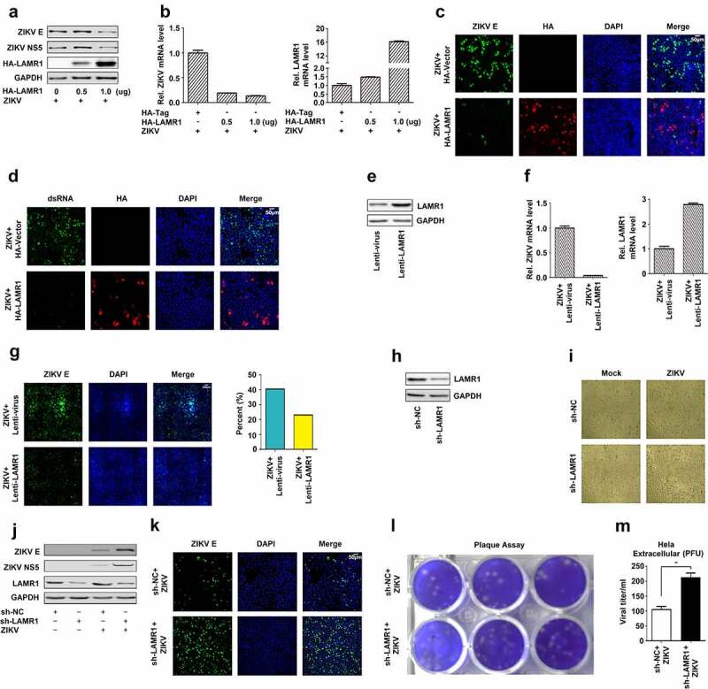
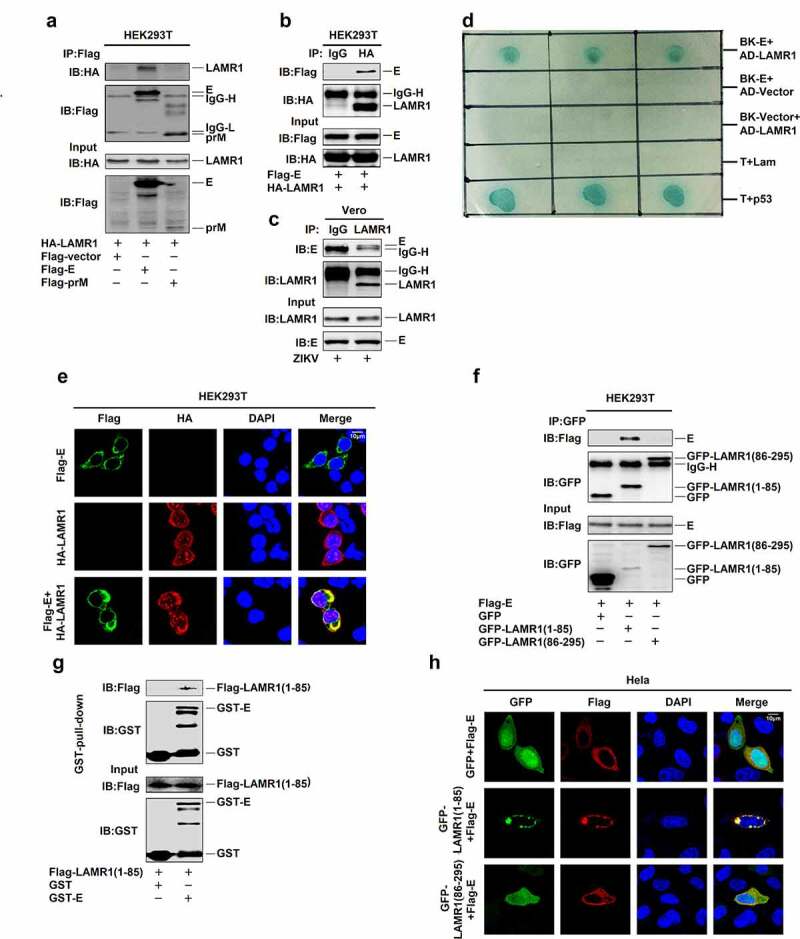
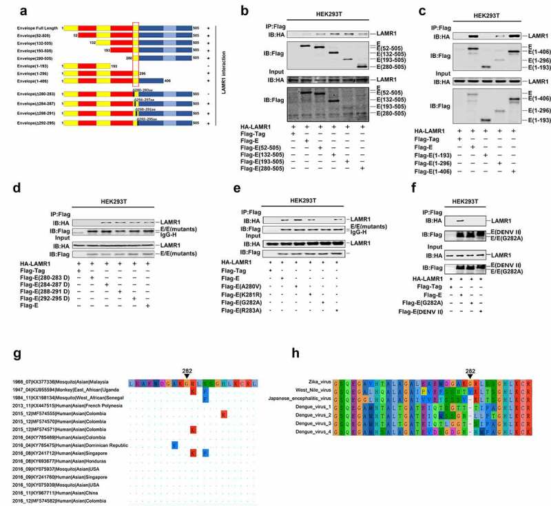
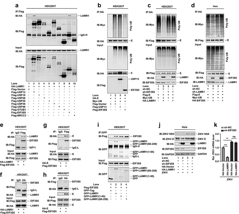
Zika virus (ZIKV) infection can cause severe neurological disorders, including Guillain-Barre syndrome and meningoencephalitis in adults and microcephaly in fetuses. Here, we reveal that laminin receptor 1 (LAMR1) is a novel host resistance factor against ZIKV infection. Mechanistically, we found that LAMR1 binds to ZIKV envelope (E) protein via its intracellular region and attenuates E protein ubiquitination through recruiting the deubiquitinase eukaryotic translation initiation factor 3 subunit 5 (EIF3S5). We further found that the conserved G282 residue of E protein is essential for its interaction with LAMR1. Moreover, a G282A substitution abolished the binding of E protein to LAMR1 and inhibited LAMR1-mediated E protein deubiquitination. Together, our results indicated that LAMR1 represses ZIKV infection through binding to E protein and attenuating its ubiquitination.

Keywords: E protein; Laminin receptor 1, LAMR1; Zika virus, ZIKV; eukaryotic translation initiation factor 3 subunit 5, EIF3S5; ubiquitination.


Conflict of interest statement

No potential conflict of interest was reported by the authors.

Figures

Figure 1.
Figure 1.
LAMR1 is a host restriction factor against ZIKV infection. (a–d) HeLa cells were transfected with pHA-LAMR1 or empty vector for 16 h and then infected with ZIKV (MOI = 1) for 48 h. The expression levels of ZIKV proteins were detected by immunoblotting (a) and confocal microscopy (c) and the viral RNA content was quantified by qPCR (b) and confocal microscopy (d). (e–g) Hela cells stably expressing LAMR1 or the control gene were generated and analyzed(e). Cells were infected with ZIKV (MOI = 1) for 48 h, following which viral RNA levels were quantified by qPCR (f) and ZIKV E protein levels by high-content analysis (g). (h–m) HeLa cells stably expressing sh-LAMR1 or control sh-RNA were generated and analyzed(h). Cells were infected with ZIKV (MOI = 1) for 48 h, and the cytopathic effects of cells was captured under the microscope (i). The expression levels of viral proteins were assessed by immunoblotting (j), while ZIKV titer in supernatants was calculated through a plaque assay (l, m)
Figure 2.
Figure 2.
LAMR1 binds to ZIKV E protein through its intracellular domain. (a) HEK293T cells were transfected with plasmids encoding HA-LAMR1 and Flag-E or pFlag-prM. Cell lystes were prepared using lysis buffer and then used for immunoprecipitation (IP) with anti-Flag antibody and analyzed by SDS-PAGE. (b) HEK293T cells were transfected with plasmids encoding HA-LAMR1 and Flag-E. Cell lysates were prepared using lysis buffer and then used for IP with anti-Flag antibody or control IgG and analyzed by SDS-PAGE. (c) Vero cells were infected with ZIKV (MOI = 1) for 48 h and then subjected to IP with anti-LAMR1 antibody or control IgG. (d) A yeast two-hybrid screen was used to identify the interaction between LAMR1 and E protein. (e) HEK293T cells were transfected with pFlag-E or pHA-LAMR1, or co-transfected with pFlag-E and pHA-LAMR1. Immunofluorescence staining showed the sub-cellular localization of, HA-LAMR1 (red) and Flag-E (green); the nucleus is marked with DAPI (blue). (f) HEK293T cells were transfected with plasmids encoding Flag-E and GFP-vector/GFP-LAMR1 (1–85)/GFP-LAMR1 (86–295). Cell lysates were prepared using lysis buffer and then used for IP with anti-GFP antibody and analyzed by SDS-PAGE. (g) The Glutathione Sepharose beads were added to GST-E protein or GST only. The mixtures were then incubated with whole-cell extracts of HEK293T cells transfected with a plasmid encoding Flag-LAMR1 (1–85) (h) HEK293T cells were transfected with pFlag-E and pGFP-vector, pGFP-LAMR1 (1–85), and pGFP-LAMR1 (86–295). The sub-cellular localization of GFP, GFP-LAMR1 (1–85), GFP-LAMR1 (86–295) (green) and Flag-E (red) were analyzed by confocal microscopy; the nucleus is marked with DAPI (blue)
Figure 3.
Figure 3.
The conserved E protein G282 residue is essential for the binding of LAMR1. (a) Diagrams of the full-length E protein, truncated forms of E protein (1–505, 52–505, 132–505, 193–505, 280–505, 1–93, 1–296, and 1–406) and deletion forms of E protein (Δ280–283, Δ284–287, Δ288–291, and Δ292–295). (b) HEK293T cells were transfected with plasmids encoding HA-LAMR1 and Flag-vector, Flag-E/Flag-E (52–505), Flag-E (132–505), Flag-E (193–505), and Flag-E (280–505). Cell lysates were prepared using lysis buffer and then analyzed by immunoprecipitation with the indicated antibodies. (c) HEK293T cells were transfected with plasmids encoding HA-LAMR1 and pFlag-vector, Flag-E/Flag-E (1–193), Flag-E (1–296), and pFlag-E (1–406). Cell lysates were prepared using lysis buffer and then analyzed by immunoprecipitation with the indicated antibodies. (d) HEK293T cells were transfected with plasmids encoding HA-LAMR1 and Flag-vector, Flag-E, Flag-E (280–283aa deletion), Flag-E (284–287aa deletion), Flag-E (288–291aa deletion), and Flag-E (292–295aa deletion). Cell lysates were prepared using lysis buffer and then analyzed by immunoprecipitation with the indicated antibodies. (e) HEK293T cells were transfected with plasmids encoding HA-LAMR1 and Flag-vector, Flag-E, Flag-E G282A, and Flag-E DENV II. Cell lysates were prepared with lysis buffer and then analyzed by immunoprecipitation with indicated antibodies. (f) HEK293T cells were transfected with plasmids encoding HA-LAMR1 and Flag-vector, Flag-E, Flag-E A280V, Flag-E K281R, Flag-E G282A, and Flag-E R283A. Cell lysates were prepared with lysis buffer and then analyzed by immunoprecipitation with indicated antibodies. (g) Diagram of the consered G282 site. Viral sequences were downloaded from GenBank, and viewed and aligned using AliView software. (h) The diagrams of amino acid sequences of partial E protein among ZIKV, WNV, JEV and DENV. Sequences were downloaded from GenBank and viewed and aligned using AliView software
Figure 4.
Figure 4.
LAMR1 attenuates K48 – and K63-linked E protein polyubiquitination. (a, b) HEK293T cells and HeLa cells were co-transfected with pFlag-E, pMyc-UB, and pHA-LAMR1. Lysates were prepared and used for IP with the indicated antibodies and analyzed by SDS-PAGE. (c) HEK293T cells were transfected with pFlag-E, pMyc-UB and GFP, GFP-LAMR1, GFP-LAMR1 (1–85) and GFP-LAMR1 (86–295). Lysates were prepared and used for IP with the indicated antibodies and analyzed by SDS-PAGE. (d, e) HEK293T cells were co-transfected with pFlag-E, pMyc-UB, pMyc-UB K48O, pMyc-UB K63O, pMyc-UB K48R, Myc-UB K63R and HA-LAMR1. Lysates were prepared and used for IP with indicated antibodies and analyzed by SDS-PAGE. (f, g) HEK293T cells were co-transfected with pFlag-E, pFlag-E K38R, pFlag-E K281R, pFlag-E K38&281R, pFlag-E A280V, pFlag-E G282A, pFlag-E R283A, Myc-UB and HA-LAMR1. Lysates were prepared and used for IP with the indicated antibodies and analyzed by SDS-PAGE
Figure 5.
Figure 5.
LAMR1 recruits EIF3S5 to deubiquitinate ZIKV E protein. (a) HEK293T cells were co-transfected with pHA-LAMR1 and pFlag-USP13, pFlag-USP15, pFlag-USP26, pFlag-USP30, pFlag-USP38, pFlag-USP49, pFlag-OTUB1, pFlag-EIF3S5, or pFlag-BRCC3. Lysates were prepared and used for IP with an anti-Flag antibody and analyzed by SDS-PAGE. (b) HEK293T cells were co-transfected with pHA-E, pMyc-UB, pFlag-USP13, and pFlag-EIF3S5. Lysates were prepared and used for IP with an anti-HA antibody and then analyzed by SDS-PAGE. (c) EIF3S5-knockdown HEK293T cells and control cells were co-transfected with pFlag-E, pMyc-UB, and pHA-LAMR1. Lysates were prepared and used for IP with an anti-Flag antibody and then analyzed by SDS-PAGE. (d) LAMR1-knockdown HeLa cells and control cells were co-transfected with pFlag-E, pMyc-UB, and pHA-EIF3S5. Lysates were prepared and used for IP with an anti-Flag antibody and then analyzed by SDS-PAGE. (e, f) HEK293T cells were transfected with plasmids encoding Flag-EIF3S5 and HA-LAMR1. Cell lysates were prepared with lysis buffer and then analyzed by IP with the indicated antibodies. (g, h) HEK293T cells were transfected with plasmids encoding Flag-EIF3S5 and HA-E. Cell lysates were prepared with lysis buffer and then analyzed by IP with indicated antibodies and immunoblotting as described above. (i) HEK293T cells were transfected with plasmids encoding Flag-EIF3S5 and GFP-LAMR1, GFP-LAMR1 (1–85aa), and GFP-LAMR1 (86–259aa). Cell lysates were prepared with lysis buffer and then analyzed by IP with the indicated antibodies. (j, k) HeLa cells stably expressing sh-EIF3S5 or control sh-RNA were generated and analyzed. Cells were transfected with pHA-LAMR1 or empty vector for 16 h, and then infected with ZIKV (MOI = 1) for 48 h. The levels of viral protein and RNA were detected by immunoblotting and qPCR, respectively

Similar articles

Cited by

References

    1. Musso D, Gubler DJ.. Zika virus. Clin Microbiol Rev. 2016;29(3):487–524. - PMC - PubMed
    1. Dick GW, Kitchen SF, HADDOW AJ, et al. Isolations and serological specificity. Trans R Soc Trop Med Hyg. 1952;46(5):509–520. - PubMed
    1. Duffy MR, Chen TH, Hancock WT, et al. Zika virus outbreak on yap island, federated states of micronesia. N Engl J Med. 2009;360(24):2536–2543. - PubMed
    1. Faria NR, Azevedo R, Kraemer M, et al. Zika virus in the Americas: Early epidemiological and genetic findings. Science. 2016;352(6283):345–349. - PMC - PubMed
    1. Pierson TC, Diamond MS. The emergence of Zika virus and its new clinical syndromes. Nature. 2018;560(7720):573–581. - PubMed

Publication types