Review
. 2021 Aug 27;49(4):1695-1709.
doi: 10.1042/BST20201144.

Emerging roles of adhesion G protein-coupled receptors

Affiliations
Review

Emerging roles of adhesion G protein-coupled receptors

Matthew Rosa et al. Biochem Soc Trans. .

Abstract

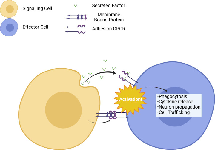
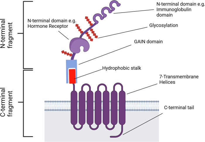
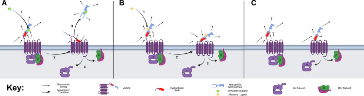
Adhesion G protein-coupled receptors (aGPCRs) form a sub-group within the GPCR superfamily. Their distinctive structure contains an abnormally large N-terminal, extracellular region with a GPCR autoproteolysis-inducing (GAIN) domain. In most aGPCRs, the GAIN domain constitutively cleaves the receptor into two fragments. This process is often required for aGPCR signalling. Over the last two decades, much research has focussed on aGPCR-ligand interactions, in an attempt to deorphanize the family. Most ligands have been found to bind to regions N-terminal to the GAIN domain. These receptors may bind a variety of ligands, ranging across membrane-bound proteins and extracellular matrix components. Recent advancements have revealed a conserved method of aGPCR activation involving a tethered ligand within the GAIN domain. Evidence for this comes from increased activity in receptor mutants exposing the tethered ligand. As a result, G protein-coupling partners of aGPCRs have been more extensively characterised, making use of their tethered ligand to create constitutively active mutants. This has led to demonstrations of aGPCR function in, for example, neurodevelopment and tumour growth. However, questions remain around the ligands that may bind many aGPCRs, how this binding is translated into changes in the GAIN domain, and the exact mechanism of aGPCR activation following GAIN domain conformational changes. This review aims to examine the current knowledge around aGPCR activation, including ligand binding sites, the mechanism of GAIN domain-mediated receptor activation and how aGPCR transmembrane domains may relate to activation. Other aspects of aGPCR signalling will be touched upon, such as downstream effectors and physiological roles.

Keywords: G-protein-coupled receptors; G-proteins; adhesion receptors; agonists; signal transduction.


Conflict of interest statement

The authors declare that there are no competing interests associated with the manuscript.

Figures

Figure 1.
Figure 1.. Types of signalling between cells using aGPCRs.
aGPCRs are mainly utilised in paracrine or autocrine signalling via either secreted factors (top) or membrane-bound proteins and proteoglycans on adjacent cells (bottom). Activation through either of these two methods can lead to a cellular response. Created with Biorender.
Figure 2.
Figure 2.. Example aGPCR structure.
The GPS, dividing the N- and C-terminal fragments, lies between the hydrophobic stalk and GAIN domain. Created with Biorender.
Figure 3.
Figure 3.. Proposed activation states of aGPCRs and the corresponding electrostatic forces.
Inactive aGPCRs have their G proteins bound and stalks away from the activation domain in the centre of the GPCR. This is due to the hydrophilic GAIN domain still being attached and the hydrophobic stalk being hidden within it. (A) Full activation of the aGPCR is achieved by autoproteolysis of the GAIN domain, to expose the hydrophobic stalk to the ECM, pushing it toward the hydrophobic centre of the activation domain. This activates the GPCR releasing the G protein causing further downstream effects. (B) Partial allosteric activation can result in a conformational change of the GAIN domain resulting in the exposure of part of the hydrophobic stalk. This pushes the stalk toward the activation domain resulting in a higher chance of the G protein subunit dissociating. (C) Some receptors have constitutive activity, and this is likely due to the exposure of some of the hydrophobic residues on the stalk, resulting in more forces pushing the stalk away from the water rich ECM and toward the hydrophobic centre of the aGPCR. This can partially activate the aGPCR resulting in a higher chance of G protein subunit dissociation and downstream effects. Created using Biorender.

References

    1. Sriram, K. and Insel, P.A. (2018) G protein-coupled receptors as targets for approved drugs: how many targets and how many drugs? Mol. Pharmacol. 93, 251–258 10.1124/mol.117.111062 - DOI - PMC - PubMed
    1. Lagerström, M.C. and Schiöth, H.B. (2008) Structural diversity of G protein-coupled receptors and significance for drug discovery. Nat. Rev. Drug Discov. 7, 339–357 10.1038/nrd2518 - DOI - PubMed
    1. Bondarev, A.D., Attwood, M.M., Jonsson, J., Chubarev, V.N., Tarasov, V.V. and Schiöth, H.B. (2020) Opportunities and challenges for drug discovery in modulating adhesion G protein-coupled receptor (GPCR) functions. Expert Opin. Drug Discov. 15, 1291–1307 10.1080/17460441.2020.1791075 - DOI - PubMed
    1. Yona, S., Lin, H.-H., Siu, W.O., Gordon, S. and Stacey, M. (2008) Adhesion-GPCRs: emerging roles for novel receptors. Trends Biochem. Sci. 33, 491–500 10.1016/j.tibs.2008.07.005 - DOI - PubMed
    1. Gad, A.A. and Balenga, N. (2020) The emerging role of adhesion GPCRs in cancer. ACS Pharmacol. Transl. Sci. 3, 29–42 10.1021/acsptsci.9b00093 - DOI - PMC - PubMed

Publication types