Review
. 2021 Jul 2;13(13):3328.
doi: 10.3390/cancers13133328.

Adipose Tissue-Derived Extracellular Vesicles and the Tumor Microenvironment: Revisiting the Hallmarks of Cancer

Affiliations
Review

Adipose Tissue-Derived Extracellular Vesicles and the Tumor Microenvironment: Revisiting the Hallmarks of Cancer

João Alfredo Moraes et al. Cancers (Basel). .

Abstract

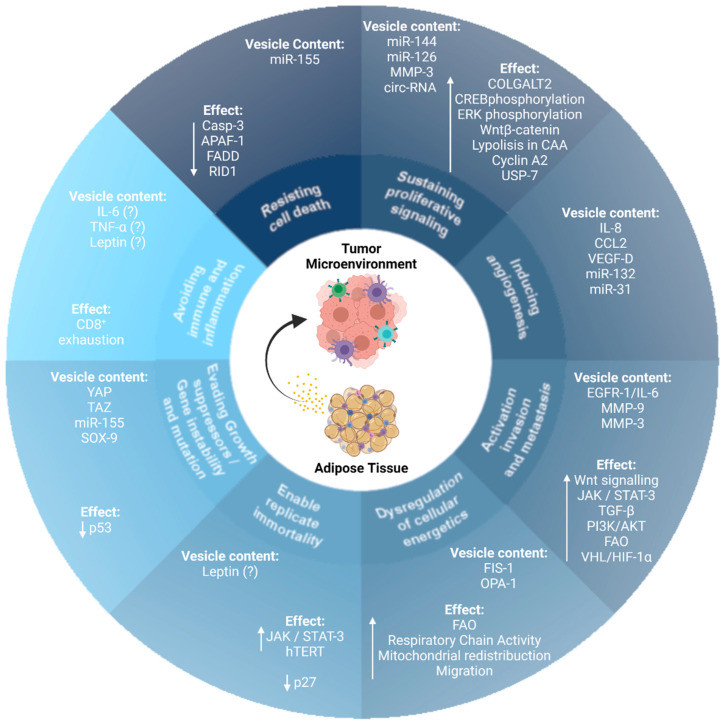
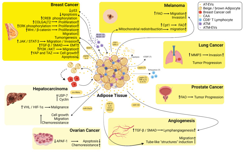
Extracellular vesicles (EVs) are crucial elements that sustain the communication between tumor cells and their microenvironment, and have emerged as a widespread mechanism of tumor formation and metastasis. In obesity, the adipose tissue becomes hypertrophic and hyperplastic, triggering increased production of pro-inflammatory adipokines, such as tumor necrosis factor α, interleukin 6, interleukin 1, and leptin. Furthermore, obese adipose tissue undergoes dysregulation in the cargo content of the released EVs, resulting in an increased content of pro-inflammatory proteins, fatty acids, and oncogenic microRNAs. These alterations drive obesity-associated inflammatory responses both locally and systemically. After being ignored for a long time, adipose tissues have recently received considerable attention as a major player in tumor microenvironment-linked obesity and cancer. The role of adipose tissue in the establishment and progression of cancer is reinforced by its high plasticity and inflammatory content. Such a relationship may be established by direct contact between adipocytes and cancer cells within the microenvironment or systemically, via EV-mediated cell-to-cell communication. Here, we highlight cues evidencing the influence of adipose tissue-derived EVs on the hallmarks of cancer, which are critical for tumor malignancy.

Keywords: adipose tissue; cancer; extracellular vesicles; hallmarks of cancer; tumor microenvironment.


Conflict of interest statement

The authors declare that they have no conflict of interest.

Figures

Figure 1
Figure 1
Extracellular vesicles (EVs) derived from adipose tissues modulate the acquisition and maintenance of cancer hallmark traits. EVs derived from adipose tissue modulate the acquisition and maintenance of the hallmark traits of cancer. MicroRNA (miR); circulating-RNA (circ-RNA); procollagen galactosyltransferase 2 (COLGALT2); cAMP response element-binding protein (CREB); CAA (cancer-associated adipocytes); ubiquitin-specific-processing protease 7 (USP-7); interleukin-8 (IL-8), C-C motif chemokine ligand 2 (CCL2), and vascular endothelial growth factor-D (VEGF-D); epidermal growth factor receptor 1 (EGFR1); interleukin 6 (IL-6); metalloproteinase 3 (MMP-3); metalloproteinase 9 (MMP-9); transforming growth factor β (TGF-β); janus kinase (JAK); signal transducer and activator of transcription 3 (STAT-3); hypoxia-inducible factor-1 α (HIF1-α); von-Hippel Lindau tumor suppressor (VHL); mitochondrial fission protein 1 (FIS-1); mitochondrial dynamin-like GTPase (OPA-1); fatty acid oxidation (FAO); human telomerase reverse transcriptase (hTERT); yes-associated protein 1 (YAP); taffazin (TAZ); caspase 3 (Casp-3); apoptotic peptidase activator factor 1 (APAF-1); Fas-associated death domain protein (FADD); GTPase-binding protein rid1 (RID1); SRY-box transcription factor 9 (SOX-9).
Figure 2
Figure 2
Summary of the crosstalk between the EVs derived from adipose tissues (AT-EVs) and several types of cancers. EVs released from adipose tissue macrophages are enriched in miR-155, whereas miR-144 is downregulated inducing inhibition of p53 and decreased apoptosis in breast cancer cells. AT-EVs increase phosphorylation of cAMP response element-binding (CREB) protein increasing the proliferation of breast cancer cells. AT-EVs increase the proliferation of breast cancer cells via induction of procollagen galactosyltransferase 2 (COLGALT2). AT-EVs increase extracellular signal-regulated kinase (ERK) and Wnt/β-catenin signaling pathways inducing an increase in the proliferation of breast cancer cells. AT-EVs enriched in SRY-box transcription factor 9 (SOX-9) induce tumorigenesis in breast cancer cells. AT-EVs from diabetic patients are enriched in epidermal growth factor receptor 1 (EGFR1)/interleukin 6 (IL-6) and induce an increase in migration and invasion of breast cancer cells via Janus kinase (JAK)/signal transducer and activator of transcription 3 (STAT-3) signaling. AT-EVs increase TGF-β/SMAD signaling pathway triggering epithelial-mesenchymal transition (EMT) in non-metastatic breast cancer cells. AT-EVs induce PI3K/AKT signaling in MDA-MB-231 cells, thus increasing their migratory capacity. AT-EVs induce yes-associated protein 1 (YAP) and taffazin (TAZ) inducing cell growth in breast cancer cells. AT-EVs decrease apoptosis in breast cancer cells. AT-EVs enriched in mitochondrial fission protein 1 (FIS-1) and mitochondrial dynamin-like GTPase (OPA-1) induce mitochondrial redistribution toward the edge of melanoma cells increasing their migratory capacity. AT-EVs and EVs derived from cancer-associated adipocytes (CAA) fuel free fatty acids to melanoma cells and increase fatty acid oxidation (FAO) increasing their migratory and invasive capacities. AT-EVs increase metalloproteinase 3 (MMP-3) in lung cancer cells increasing their invasiveness. AT-EVs increase tumor progression of lung cancer. AT-EVs increase FAO and induce tumor progression in prostate cancer. AT-EVs enriched in circulating RNAs (circ-RNAs) induce hypophosphorylation of ubiquitin-specific-processing protease 7 (USP-7) and an increase in cyclin in hepatocarcinoma cells. AT-EVs induce malignancy of hepatocarcinoma cells via hypoxia-inducible factor 1 α (HIF-1α) binding to the von-Hippel Lindau tumor suppressor (VHL). AT-EVs decrease apoptosis and increase chemoresistance in ovarian cancer cells via inhibition of the apoptotic peptidase activator factor 1 (APAF-1). AT-EVs enriched in miR-31, interleukin 8 (IL-8), C-C motif chemokine ligand 2 (CCL2), and vascular endothelial growth factor-D (VEGF-D) induce the formation of tube-like structures by endothelial cells. AT-EVs enriched in miR-132 induce lymphangiogenesis via transforming growth factor β (TGF-β)/Smad signaling. CAA undergo increased lipolysis and differentiate toward beige/brown adipocytes phenotype fueling tumor cells with EVs. Leptin released from AT is possibly within EVs and induces an increase in telomerase reverse transcriptase (hTERT) activity inducing resistance to apoptosis and senescence. Leptin, TNF-α, and IL-6 may be released from AT-EVs, inducing CD8+ T cells’ exhaustion.

Similar articles

Cited by

References

    1. Avgerinos K.I., Spyrou N., Mantzoros C.S., Dalamaga M. Obesity and cancer risk: Emerging biological mechanisms and perspectives. Metabolism. 2019;92:121–135. doi: 10.1016/j.metabol.2018.11.001. - DOI - PubMed
    1. Booth A., Magnuson A., Fouts J., Foster M. Adipose tissue, obesity and adipokines: Role in cancer promotion. Horm. Mol. Biol. Clin. Investig. 2015;21:57–74. doi: 10.1515/hmbci-2014-0037. - DOI - PubMed
    1. Steele C.B., Thomas C.C., Henley J., Massetti G.M., Galuska D.A., Agurs-Collins T., Puckett M., Richardson L.C. Vital Signs: Trends in Incidence of Cancers Associated with Overweight and Obesity—United States, 2005–2014. MMWR Morb. Mortal. Wkly. Rep. 2017;66:1052–1058. doi: 10.15585/mmwr.mm6639e1. - DOI - PMC - PubMed
    1. Schauer D.P., Feigelson H.S., Koebnick C., Caan B., Weinmann S., Leonard A.C., Powers J.D., Yenumula P.R., Arterburn D.E. Association Between Weight Loss and the Risk of Cancer after Bariatric Surgery. Obesity (Silver Spring) 2017;25(Suppl. 2):S52–S57. doi: 10.1002/oby.22002. - DOI - PMC - PubMed
    1. Lauby-Secretan B., Scoccianti C., Loomis D., Grosse Y., Bianchini F., Straif K., International Agency for Research on Cancer Handbook Working Group Body Fatness and Cancer—Viewpoint of the IARC Working Group. N. Engl. J. Med. 2016;375:794–798. doi: 10.1056/NEJMsr1606602. - DOI - PMC - PubMed

LinkOut - more resources