

The Analysis of Inflammation-Related Proteins in a Cargo of Exosomes Derived from the Serum of Uveal Melanoma Patients Reveals Potential Biomarkers of Disease Progression
- PMID: 34283046
- PMCID: PMC8268237
- DOI: 10.3390/cancers13133334
The Analysis of Inflammation-Related Proteins in a Cargo of Exosomes Derived from the Serum of Uveal Melanoma Patients Reveals Potential Biomarkers of Disease Progression
Abstract
Background: Uveal melanoma (UM) is the most common intraocular tumour in adults with a poor prognosis and extremely high mortality rate due to the development of metastatic disease. However, despite relatively good knowledge about the histological and genetic risk factors for metastasis development, there is no specific biomarker that would allow early detection of UM progression. Recently, exosomes and their molecular cargo have been widely studied in the search for potential biomarkers in several cancers. The purpose of this study was to analyze the inflammation-related protein cargo of exosomes derived from the serum of primary and metastatic UM patients and healthy donors.
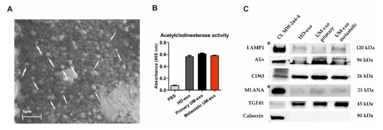
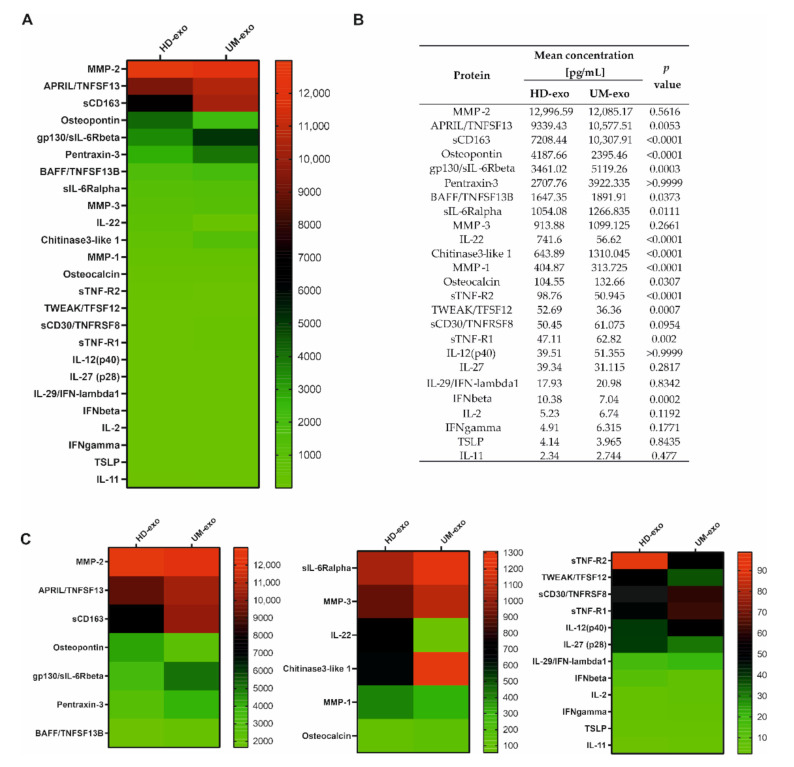
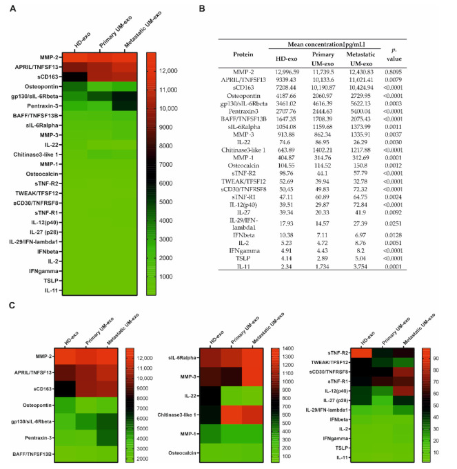
Methods: The exosomes were isolated from the serum of primary and metastatic UM patients and healthy donors. Using multiplex immunoassay technology, we analyzed the concentration of 37 inflammation-related proteins in obtained exosomes.
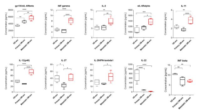
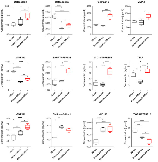
Results: The analysis of protein cargo showed several molecules related to inflammation, such as interferon-gamma, interleukin 2, 22 and 12(p40), Pentraxin-3, TNFSF13B and TNFSF8 which were significantly enriched in metastatic UM exosomes. We showed a significant correlation between the disease stage and the concentration of these inflammation-related proteins from exosomal cargo.
Conclusions: Based on the obtained results, we propose the panel of exosomal proteins for early detection of uveal melanoma progression into metastatic disease.
Keywords: biomarkers; exosomes; inflammation-related proteins; uveal melanoma.
Conflict of interest statement
The authors declare no conflict of interest.
Figures
Similar articles
-
MiRNAs from serum-derived extracellular vesicles as biomarkers for uveal melanoma progression.Front Cell Dev Biol. 2022 Dec 22;10:1008901. doi: 10.3389/fcell.2022.1008901. eCollection 2022. Front Cell Dev Biol. 2022. PMID: 36619870 Free PMC article.
-
miRNA profiling in vitreous humor, vitreal exosomes and serum from uveal melanoma patients: Pathological and diagnostic implications.Cancer Biol Ther. 2015;16(9):1387-96. doi: 10.1080/15384047.2015.1046021. Epub 2015 May 7. Cancer Biol Ther. 2015. PMID: 25951497 Free PMC article.
-
New Insights into the Exosome-Induced Migration of Uveal Melanoma Cells and the Pre-Metastatic Niche Formation in the Liver.Cancers (Basel). 2024 Aug 27;16(17):2977. doi: 10.3390/cancers16172977. Cancers (Basel). 2024. PMID: 39272836 Free PMC article.
-
MicroRNAs and Uveal Melanoma: Understanding the Diverse Role of These Small Molecular Regulators.Int J Mol Sci. 2020 Aug 6;21(16):5648. doi: 10.3390/ijms21165648. Int J Mol Sci. 2020. PMID: 32781746 Free PMC article. Review.
-
Proteomics in uveal melanoma research: opportunities and challenges in biomarker discovery.Expert Rev Proteomics. 2007 Apr;4(2):273-86. doi: 10.1586/14789450.4.2.273. Expert Rev Proteomics. 2007. PMID: 17425462 Review.
Cited by
-
Recent Advances in Molecular and Genetic Research on Uveal Melanoma.Cells. 2024 Jun 12;13(12):1023. doi: 10.3390/cells13121023. Cells. 2024. PMID: 38920653 Free PMC article. Review.
-
Dynamic interactions in the tumor niche: how the cross-talk between CAFs and the tumor microenvironment impacts resistance to therapy.Front Mol Biosci. 2024 Feb 22;11:1343523. doi: 10.3389/fmolb.2024.1343523. eCollection 2024. Front Mol Biosci. 2024. PMID: 38455762 Free PMC article. Review.
-
Exosomes are the mediators between the tumor microenvironment and prostate cancer (Review).Exp Ther Med. 2024 Sep 25;28(6):439. doi: 10.3892/etm.2024.12728. eCollection 2024 Dec. Exp Ther Med. 2024. PMID: 39355518 Free PMC article. Review.
-
Uveal Melanoma Patients Have a Distinct Metabolic Phenotype in Peripheral Blood.Int J Mol Sci. 2023 Mar 7;24(6):5077. doi: 10.3390/ijms24065077. Int J Mol Sci. 2023. PMID: 36982149 Free PMC article.
-
Opposing Effects of Chelidonine on Tyrosine and Serine Phosphorylation of STAT3 in Human Uveal Melanoma Cells.Int J Mol Sci. 2021 Nov 30;22(23):12974. doi: 10.3390/ijms222312974. Int J Mol Sci. 2021. PMID: 34884773 Free PMC article.
References
-
- Robertson A.G., Shih J., Yau C., Gibb E.A., Oba J., Mungall K.L., Hess J.M., Uzunangelov V., Walter V., Danilova L., et al. Integrative Analysis Identifies Four Molecular and Clinical Subsets in Uveal Melanoma. Cancer Cell. 2017;32:204–220.e215. doi: 10.1016/j.ccell.2017.07.003. - DOI - PMC - PubMed
Grants and funding
LinkOut - more resources
Full Text Sources
Other Literature Sources
