Review
. 2021 Jul 4;21(13):4584.
doi: 10.3390/s21134584.

Dielectric Metalens: Properties and Three-Dimensional Imaging Applications

Affiliations
Review

Dielectric Metalens: Properties and Three-Dimensional Imaging Applications

Sun-Je Kim et al. Sensors (Basel). .

Abstract

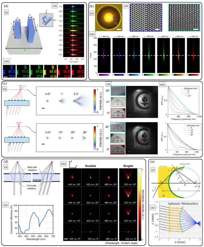
Recently, optical dielectric metasurfaces, ultrathin optical skins with densely arranged dielectric nanoantennas, have arisen as next-generation technologies with merits for miniaturization and functional improvement of conventional optical components. In particular, dielectric metalenses capable of optical focusing and imaging have attracted enormous attention from academic and industrial communities of optics. They can offer cutting-edge lensing functions owing to arbitrary wavefront encoding, polarization tunability, high efficiency, large diffraction angle, strong dispersion, and novel ultracompact integration methods. Based on the properties, dielectric metalenses have been applied to numerous three-dimensional imaging applications including wearable augmented or virtual reality displays with depth information, and optical sensing of three-dimensional position of object and various light properties. In this paper, we introduce the properties of optical dielectric metalenses, and review the working principles and recent advances in three-dimensional imaging applications based on them. The authors envision that the dielectric metalens and metasurface technologies could make breakthroughs for a wide range of compact optical systems for three-dimensional display and sensing.

Keywords: aberrations; augmented and virtual realities; depth sensing; dielectric metalens; display; flat optics; light analysis; metasurface; sensing; three-dimensional imaging.


Conflict of interest statement

The authors declare no conflict of interest.

Figures

Figure 1
Figure 1
(a) Scanning electron microscope image of the geometric-phase-based TiO2 nanofin metalens reported in 2016 [9]. (b) Optical micrograph (scale bar: 100 μm) and scanning electron microscope images of the propagation-phase-based a-Si nanopost metalens reported in 2015 [10]. The scalebars in inset electron beam images of (b) refer to 1 μm. (a) Reprinted with permission from [9]. Copyright 2016 The American Association for the Advancement of Science. (b) Reprinted with permission from [10]. Copyright 2015 Springer Nature.
Figure 2
Figure 2
Similar high numerical aperture (NA) diffraction-limited on-axis focusing performances of (a) the geometric phase based nanofin metalens by F. Capasso group [9] and (b) the propagation phase based isotropic nanopost metalens by the A. Faraon group [10]. The left sub-figure in (a) shows cross-polarization efficiency of the three types of TiO2 nanofin pixels. The central sub-figure of (a) shows measured focusing map in image space, and the right sub-figure refers to the cross-sectional point spread function at the focal point (NA: 0.8) while the collimated incident light with the wavelength of 532 nm is illuminated. In (b), transmission amplitude and phase response of the a-Si nanopost (left) according to diameter variation, point spread function at the focal point (center), and image position (d) dependent plots of focal spot size and efficiency (right), at the wavelength of 1550 nm, respectively. In the rightmost figure in (b), increase of designed d implies decrease of NA and increase of focal length. The scale bar of the central point spread function in (b) denotes 1 μm. (a) Reprinted with permission from [9]. Copyright 2016 The American Association for the Advancement of Science. (b) Reprinted with permission from [10]. Copyright 2015 Springer Nature.
Figure 3
Figure 3
(a) Schematic diagram describing the simple object imaging relation of a flat hyperbolic metalens. (b,c) Nearly diffraction-limited object imaging performances of the TiO2 nanofin metalens by Capasso group in 2016 [9]. Experimentally captured camera images of (b) 1951 United States Air Force (USAF) resolution test chart and (c) nano-scale metallic hole arrays, formed by the TiO2 nanofin metalens at the wavelength of 532 nm. The scale bars in (b) and (c) denote 40 μm, 5 μm, 10 μm, and 500 nm, from the left to right, respectively. (b,c) Reprinted with permission from [9]. Copyright 2016 The American Association for the Advancement of Science.
Figure 4
Figure 4
Performance limits of a flat hyperbolic TiO2 dielectric metalens [16]. (a) (i) Scanning electron micrographs and (ii) on-axis wavefront aberration comparison of polarization insensitive hyperbolic metalens (lower plots) with reference objective lens (upper plots) with the same NA of 0.8. The two scale bars in (i) are 5 μm (left) and 1 μm (right), respectively. In (ii), left and right plots are Zernike aberration coefficients with the Fringe notation (red: spherical aberrations) and normalized wavefront error function maps, respectively. (b) Field and wavelength-dependent focusing efficiency: comparison of the hyperbolic metalens with refractive and diffractive lenses. (i) Simulation scheme and (ii) simulated deflection efficiencies of refractive, diffractive, and metasurface lenses according to field angle. (a,b) Reprinted with permission from [16]. Copyright 2019 American Chemical Society.
Figure 5
Figure 5
(a) Longitudinal chromatic aberration correction using anisotropic complex TiO2 nanofin library [19]. (i) Nanoantenna schematic, measured broadband achromatic (ii) focusing and (iii) imaging performances. (b) Longitudinal chromatic aberration correction with GaN metalens [20]. (i) Optical image and (ii) magnified scanning electron micrographs. (iii) Measured broadband achromatic focusing property. (c) Monolithic doublet metalens with a-Si nanoposts for reducing monochromatic aberrations [25]. Comparison of singlet and doublet in terms of (i) focusing and (ii), (iii) object imaging properties. (ii) Captured sample images and corresponding (iii) modulation transfer functions for sagittal (dashed lines) and tangential (solid lines) ray directions. (d) Doublet silicon metalens optimization for simultaneous correction of chromatic and monochromatic aberrations at the three colors [26]. (i) Concept diagram, (ii) color filtering efficiency of the backside metalens, and (iii) optimized performance comparison. (e) Curved aplanatic metalens design method [27]. (i) Schematic and (ii) deterministic design without spherical aberration and coma. Reprinted with permission from (a) [19] Copyright 2018 Springer Nature, (b) [20] Copyright 2018 Springer Nature, and (c) [25] Copyright 2016 Springer Nature. Reprinted with permission from (d) [26] Copyright The Optical Society and (e) [27] Copyright The Optical Society.
Figure 6
Figure 6
Metalenses for three-dimensional display applications: near-eye displays for augmented reality (AR) and virtual reality (VR). (a) Geometric phase based metasurface eyepiece as a see-through image combiner [28]. (i) Schematic diagram and (ii) captured full-color augmented reality scene with a camera. (b) Inverse designed metalens with chromatic aberration correction and its use in near-eye virtual image generation [29]. (i) Measured longitudinal apochromatic focusing property, (ii) principle of metalens assisted fiber scanning display, and (iii) captured image of full-color augmented reality scene using a beam splitter as an image combiner. (c) Hybridization of dielectric metasurface and freeform optics [30]. (i) Ray tracing diagram of a miniature image combiner design with a metaform mirror and (ii) the imaging result of a resolution target. (iii) A possible idea to use a metaform mirror as a see-through image combiner for a wearable near-eye display for AR curved eyeglass. (a) Reprinted with permission from [28] Copyright 2018 Springer Nature. Reprinted with permission from (b) from [29] and (c) from [30] Copyright 2021 The American Association for the Advancement of Science.
Figure 7
Figure 7
(a) Spatial [34] and (b) phase [35] multiplexed light field metasurfaces for 3D sensing applications. (a) Scheme of light field imaging for depth sensing using dielectric micro-metalens array. (b) Scheme of multi-foci light field silicon metalens for real-time 3D tracking of micro-beads with enhanced resolution (left), and experimental result (right). (a) Reprinted with permission from [34] Copyright 2019 Springer Nature. (b) Reprinted with permission from [35] Copyright 2019 American Chemical Society.
Figure 8
Figure 8
Other single-shot 3D sensing technologies with dielectric metalenses. (a) Single multiplexed metalens depth sensor inspired from eye structure of jumping spider [36]. (b) Monolithic doublet metalens based quantitative phase microscopy [38]. (i) Schematic diagram and (ii) phase retrieval result of a resolution target. (iii) Accurate depth sensing result in comparison with atomic force microscopy. (c) Phase mask optimized dielectric metalens depth sensor with engineered point spread function [37]. (i) Schematic diagram and (ii) working principle. (iii) Fabrication and (iv) depth sensing results. (b) Reprinted with permission from [38] Copyright 2019 Springer Nature. (c) Reprinted with permission from [37] Copyright 2019 SPIE.
Figure 9
Figure 9
Various dielectric metalens sensing applications. (a) Micro-metalens array based polarimetric Shack-Hartmann wavefront sensor [43]. (i) Schematic diagram. Sensing results of (ii) angle of incidence and (iii) polarization map of azimuthally polarized Bessel beam. (b) Scheme of imaging polarimeter using micro-metalens array [44]. (c) Efficient quantum emitter sensing with immersion metalens [45]. (d) Folded meta-optics for compact spectrometer [46]. (e) Wavefront differentiation for edge detection using a Laplacian metasurface [47]. Fabrication result (left), edge detection of resolution target (center) and biological tissue (right). Reprinted with permission (a) from [43] Copyright 2018 Springer Nature, (b) from [44] Copyright 2018 American Chemical Society, (c) from [45] Copyright 2019 Springer Nature, (d) from [46] Copyright 2018 Springer Nature, and (e) from [47] Copyright 2020 Springer Nature.

References

    1. Chen H.T., Taylor A.J., Yu N. A review of metasurfaces: Physics and applications. Rep. Prog. Phys. 2016;79:076401. doi: 10.1088/0034-4885/79/7/076401. - DOI - PubMed
    1. Khorasaninejad M., Capasso F. Metalenses: Versatile multifunctional photonic components. Science. 2017;358:1146. doi: 10.1126/science.aam8100. - DOI - PubMed
    1. Lee Y., Kim S.J., Park H., Lee B. Metamaterials and metasurfaces for sensor applications. Sensors. 2017;17:1726. doi: 10.3390/s17081726. - DOI - PMC - PubMed
    1. Lalanne P., Chavel P. Metalenses at visible wavelengths: Past, present, perspectives. Laser Photonics Rev. 2017;11:1600295. doi: 10.1002/lpor.201600295. - DOI
    1. Lee G.-Y., Sung J., Lee B. Metasurface optics for imaging applications. MRS Bull. 2020;45:202–209. doi: 10.1557/mrs.2020.64. - DOI

LinkOut - more resources