. 2021 Jul 1;62(9):30.
doi: 10.1167/iovs.62.9.30.

Optic Nerve Engraftment of Neural Stem Cells

Affiliations

Optic Nerve Engraftment of Neural Stem Cells

Jiun L Do et al. Invest Ophthalmol Vis Sci. .

Abstract

Purpose: To evaluate the integrative potential of neural stem cells (NSCs) with the visual system and characterize effects on the survival and axonal regeneration of axotomized retinal ganglion cells (RGCs).

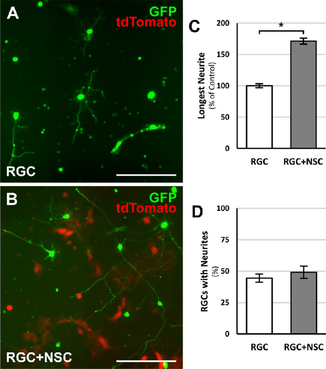
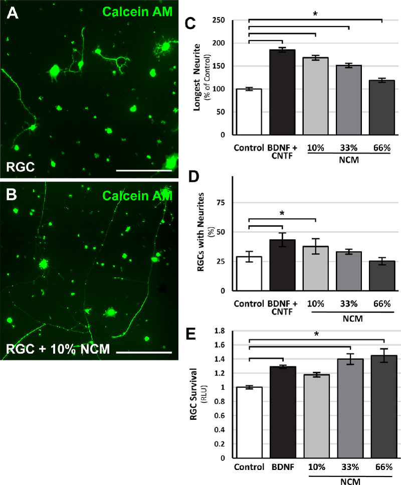
Methods: For in vitro studies, primary, postnatal rat RGCs were directly cocultured with human NSCs or cultured in NSC-conditioned media before their survival and neurite outgrowth were assessed. For in vivo studies, human NSCs were transplanted into the transected rat optic nerve, and immunohistology of the retina and optic nerve was performed to evaluate RGC survival, RGC axon regeneration, and NSC integration with the injured visual system.

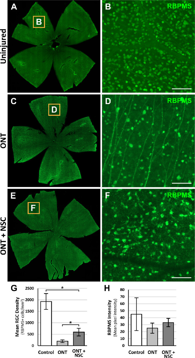
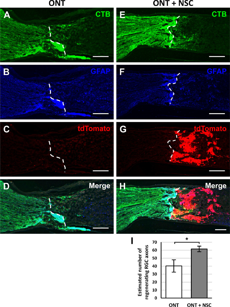
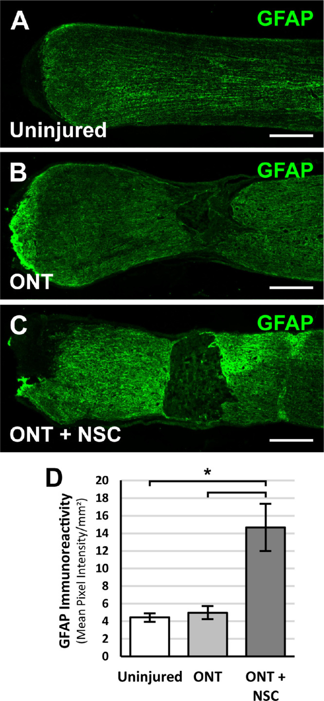
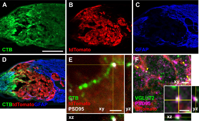
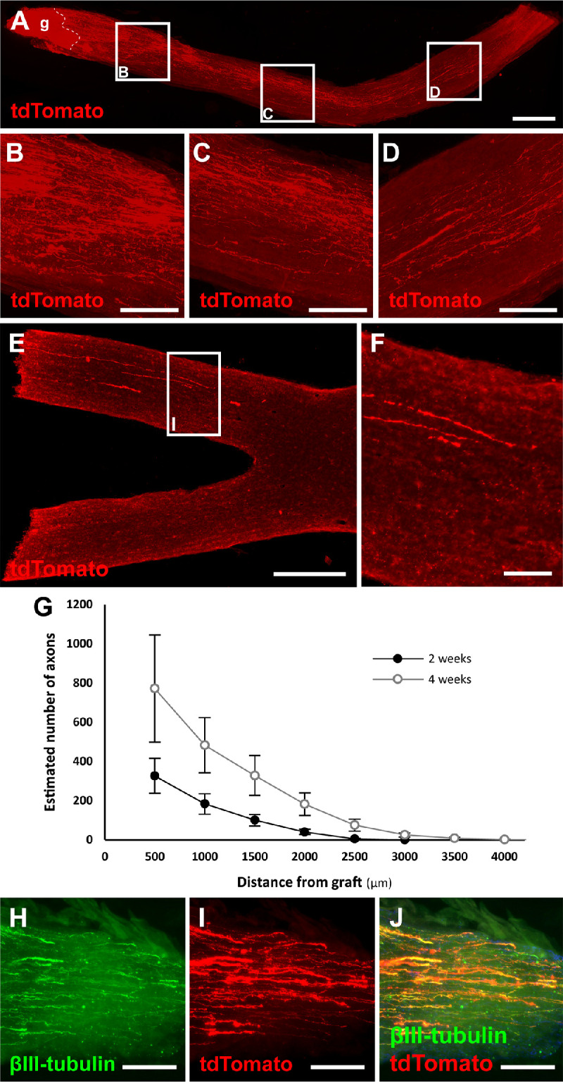
Results: Increased neurite outgrowth was observed in RGCs directly cocultured with NSCs. NSC-conditioned media demonstrated a dose-dependent effect on RGC survival and neurite outgrowth in culture. NSCs grafted into the lesioned optic nerve modestly improved RGC survival following an optic nerve transection (593 ± 164 RGCs/mm2 vs. 199 ± 58 RGCs/mm2; P < 0.01). Additionally, RGC axonal regeneration following an optic nerve transection was modestly enhanced by NSCs transplanted at the lesion site (61.6 ± 8.5 axons vs. 40.3 ± 9.1 axons, P < 0.05). Transplanted NSCs also differentiated into neurons, received synaptic inputs from regenerating RGC axons, and extended axons along the transected optic nerve to incorporate with the visual system.

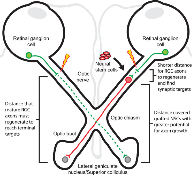
Conclusions: Human NSCs promote the modest survival and axonal regeneration of axotomized RGCs that is partially mediated by diffusible NSC-derived factors. Additionally, NSCs integrate with the injured optic nerve and have the potential to form neuronal relays to restore retinofugal connections.


Conflict of interest statement

Disclosure: J.L. Do, None; S. Allahwerdy, None; R.C.C. David, None; R.N. Weinreb, Aerie Pharmaceuticals (C), Allergan (C), Bausch & Lomb (C), Eyenovia (C), Novartis (C); Heidelberg Engineering (F), Carl Zeiss Meditec (F), Konan (F), Optovue (F), Centervue (F); M.H. Tuszynski, None; D.S. Welsbie, Oriole Therapeutics (I), Oriole Therapeutics (C), Oriole Therapeutics (P)

Figures

Figure 1.
Figure 1.
RGC-NSC direct cocultures promote RGC process outgrowth. Representative images of (A) GFP-positive RGCs cultured in isolation under standard conditions or (B) GFP-positive RGCs directly cocultured with tdTomato-expressing NSCs. (C) After two days in culture, an increase in the longest RGC process length was observed in RGCs directly cocultured with NSCs. (D) RGC-NSC direct cocultures did not change the percentage of RGCs with processes. *P < 0.05; Student's t-test. N = 4 experiments; n > 1000 cells/experiment; error bars represent SEM. Scale bars: 100 µm.
Figure 2.
Figure 2.
NSC-conditioned media promotes RGC survival and neurite outgrowth. Representative images of calcein AM-stained RGCs cultured in (A) standard RGC media or (B) 10% NSC-conditioned RGC media. RGCs cultured with BDNF and CNTF or increasing concentrations of NSC-conditioned media (NCM) demonstrated dose-dependent (C) increases in neurite outgrowth and (D) increases in the percentage of RGCs with neurite. (E) RGCs cultured with BDNF or increasing concentrations of NCM showed increase rates of survival. *P < 0.05; one-way ANOVA with post-hoc Tukey test. N = 4 experiments; n > 1000 cells/experiment; error bars represent SEM. Scale bars: 100 µm.
Figure 3.
Figure 3.
Optic nerve transplanted NSCs following ONT promote modest RGC survival. Representative retinal flat mounts stained for RBPMS and magnified insets from animals two weeks after receiving (A, B) no injury, (C, D) ONT, or (E, F) ONT with NSC graft. (G) Quantification of RGC survival demonstrates increased RGC densities with an optic nerve NSC graft compared to a ONT alone. (H) Quantification of RGC RBPMS intensity was similar among groups, suggestive of comparable cell viabilities. *P < 0.05; one-way ANOVA with post-hoc Tukey test. n = 6 animals per group; error bars represent SD. Scale bars: 100 µm. ONT, optic nerve transection.
Figure 4.
Figure 4.
Optic nerve transplanted NSCs following ONT promote modest, short-distance RGC axon regeneration. Representative images from longitudinal optic nerve sections that received an (A–D) optic nerve transection (ONT) or (E–H) an ONT with NSC graft. Dashed white lines demarcate the areas in which CTB labeling was quantified. (A, E) Intravitreal CTB injections label regenerated RGCs axons at the transection site. (B, F) Immunostaining for GFAP identifies the host tissue and was used to demarcate the proximal border of the transection (white line dotted). (C, D) NSCs constitutively express tdTomato to identify the location and extent of the cell graft. (D, H) Merged images demonstrate the localization of CTB-labeled axons, tdTomato NSCs, and GFAP borders. (I) Quantification of CTB-positive RGC axons beyond the proximal lesion border (dashed line) showed a modest increase in regenerating RGC axons when a NSC graft was present. *P < 0.05; Student's t-test. n = 6 animals per group; error bars represent SD. Scale bars: 200 µm. ONT, optic nerve transection.
Figure 5.
Figure 5.
Optic nerve transplanted NSCs following ONT modify the host response following injury. Representative images from longitudinal optic nerve sections stained for GFAP 2 weeks after (A) unlesioned control, (B) ONT, or (C) ONT with NSC graft. (D) Quantification of GFAP immunoreactivity demonstrates increased GFAP immunoreactivity at the proximal transected optic nerve end with an NSC graft. P < 0.05; one-way ANOVA with post-hoc Tukey test. n = 6 animals per group; error bars represent SD. Scale bars: 200 µm. ONT, optic nerve transection.
Figure 6.
Figure 6.
Optic nerve transplanted NSCs form synapses with transected RGC axons. (A–D) Longitudinal optic nerve section 2 weeks after optic nerve transection and grafting of tdTomato-expressing NSCs in the injury site. (A) Cholera toxin B (CTB) labeled RGC axons regenerate into the injury site. (B) NSCs expressing tdTomato transplanted into the transected optic nerve survive at the transection site. (C) GFAP labeling demarcates the glial response to injury and the boundaries of the optic nerve transection. (D) Merged images demonstrate CTB labeled RGC axons regenerating into the NSC graft. (E) Confocal image of a CTB-labeled RGC axon within the tdTomato-expressing NSC graft in proximity to the postsynaptic marker PSD-95, suggestive of synapses within the NSC graft. (F) Confocal image of VGLUT2 and PSD95 immunostaining within the tdTomato-expressing NSC graft demonstrating overlap of presynaptic and postsynaptic markers (yellow arrowheads); (F inset) high magnification of a puncta of VGLUT2 and PSD95 overlap. Scale bar: 200 µm (A–D); 2 µm (E, F); 1 µm (F inset).
Figure 7.
Figure 7.
NSCs transplanted into an optic nerve transection survive, differentiate, and extend axons toward the optic chiasm after 4 weeks. (A) Longitudinal optic nerve section 4 weeks after transection (dashed line) and tdTomato expressing NSC graft (g) extending axons along the distal optic nerve. (B–D) Magnifications of insets from (A) demonstrate robust axonal extension along the length of the optic nerve. (E, F) After only four weeks, NSC-derived axons can be seen approaching the optic chiasm. (G) Quantification of the number of NSC graft-derived axons counted at specific distances from the graft site. n = 6 animals per group; error bars represent SEM. (H–J) NSC-derived processes colocalize with the neuronal marker βIII-tubulin. Scale bar: 500 µm (A); 100 µm (B–G); 500 µm (H); 100 µm (I).
Figure 8.
Figure 8.
Schematic diagram of a NSC-derived optic nerve relay. (Left) Mature RGCs have a limited potential for axonal regeneration and, if axotomized, must regenerate a considerable distance (green dashed line) to reach targets in the lateral geniculate nucleus or superior colliculus. (Right) NSCs have a greater intrinsic potential for axon growth. When transplanted into the optic nerve to form an optic nerve relay, NSCs shorten the distance mature RGCs need to regenerate to reform synapses (green dashed line) and address the need for long-distance growth to reach terminal targets (red solid line).

References

    1. Goldberg JL, Espinosa JS, Xu Y, Davidson N, Kovacs GTA, Barres BA. Retinal ganglion cells do not extend axons by default: promotion by neurotrophic signaling and electrical activity. Neuron. 2002; 33: 689–702. - PubMed
    1. Welsbie DS, Mitchell KL, Jaskula-Ranga V, et al. .. Enhanced functional genomic screening identifies novel mediators of dual leucine zipper kinase-dependent injury signaling in neurons. Neuron. 2017; 94: 1142–1154.e6. - PMC - PubMed
    1. Bull ND, Johnson TV, Welsapar G, DeKorver NW, Tomarev SI, Martin KR.. Use of an adult rat retinal explant model for screening of potential retinal ganglion cell neuroprotective therapies. Investig Ophthalmol Vis Sci. 2011; 52: 3309–3320. - PMC - PubMed
    1. Johnson T V., Dekorver NW, Levasseur VA, et al. .. Identification of retinal ganglion cell neuroprotection conferred by platelet-derived growth factor through analysis of the mesenchymal stem cell secretome. Brain. 2014; 137: 503–519. - PMC - PubMed
    1. Lu P, Jones LL, Snyder EY, Tuszynski MH.. Neural stem cells constitutively secrete neurotrophic factors and promote extensive host axonal growth after spinal cord injury. Exp Neurol. 2003; 181: 115–129. - PubMed

Publication types