. 2021 Jul 1;62(9):27.
doi: 10.1167/iovs.62.9.27.

The Role of Retinal Connexins Cx36 and Horizontal Cell Coupling in Emmetropization in Guinea Pigs

Affiliations

The Role of Retinal Connexins Cx36 and Horizontal Cell Coupling in Emmetropization in Guinea Pigs

Zhina Zhi et al. Invest Ophthalmol Vis Sci. .

Abstract

Purpose: The purpose of this study was to determine whether retinal gap junctions (GJs) via connexin 36 (Cx36, mediating coupling of many retinal cell types) and horizontal cell (HC-HC) coupling, are involved in emmetropization.

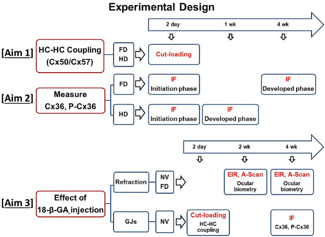
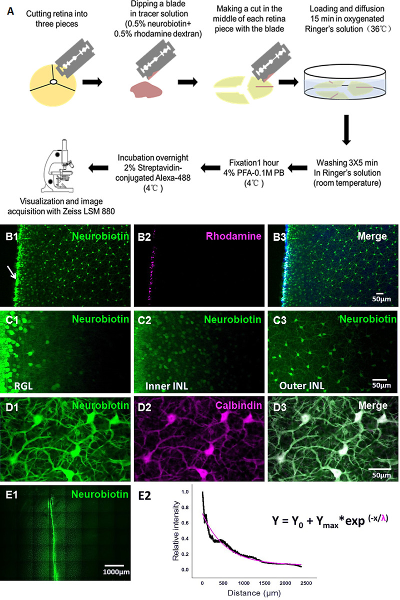
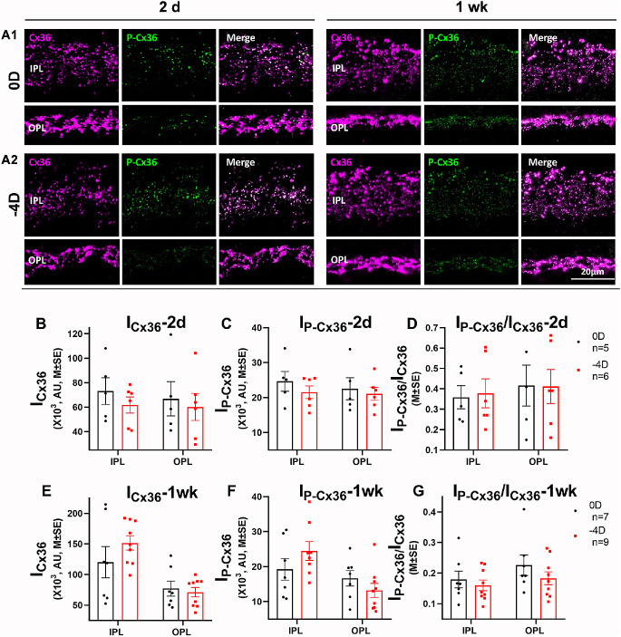
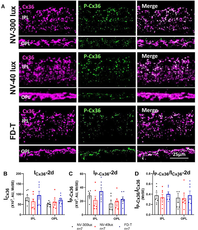
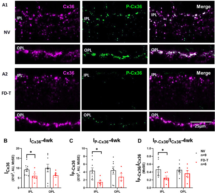
Methods: Guinea pigs (3 weeks old) were monocularly form deprived (FD) or raised without FD (in normal visual [NV] environment) for 2 days or 4 weeks; alternatively, they wore a -4 D lens (hyperopic defocus [HD]) or 0 D lens for 2 days or 1 week. FD and NV eyes received daily subconjunctival injections of a nonspecific GJ-uncoupling agent, 18-β-Glycyrrhetinic Acid (18-β-GA). The amounts of total Cx36 and of phosphorylated Cx36 (P-Cx36; activated state that increases cell-cell coupling), in the inner and outer plexiform layers (IPLs and OPLs), were evaluated by quantitative immunofluorescence (IF), and HC-HC coupling was evaluated by cut-loading with neurobiotin.

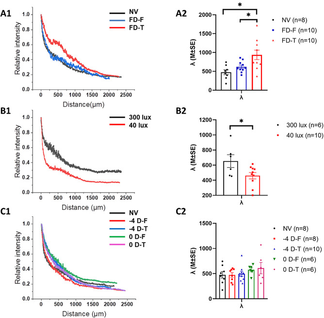
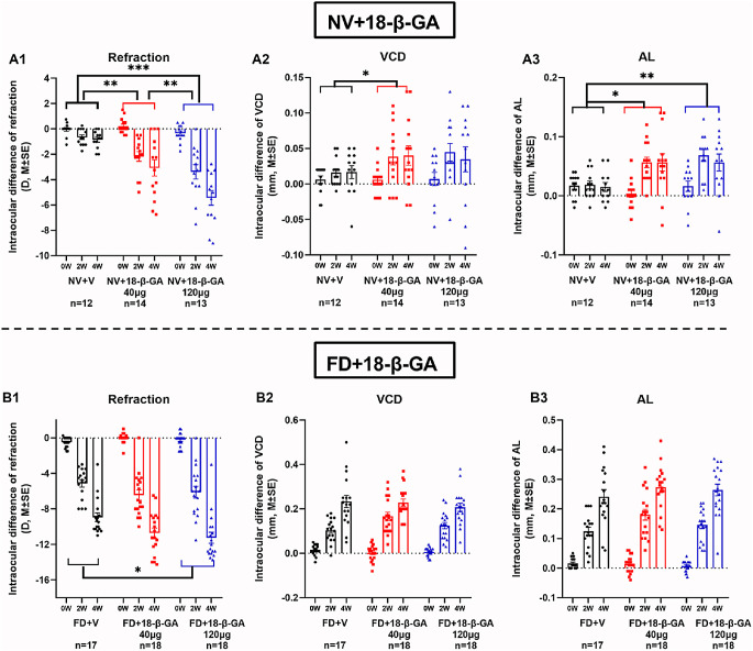
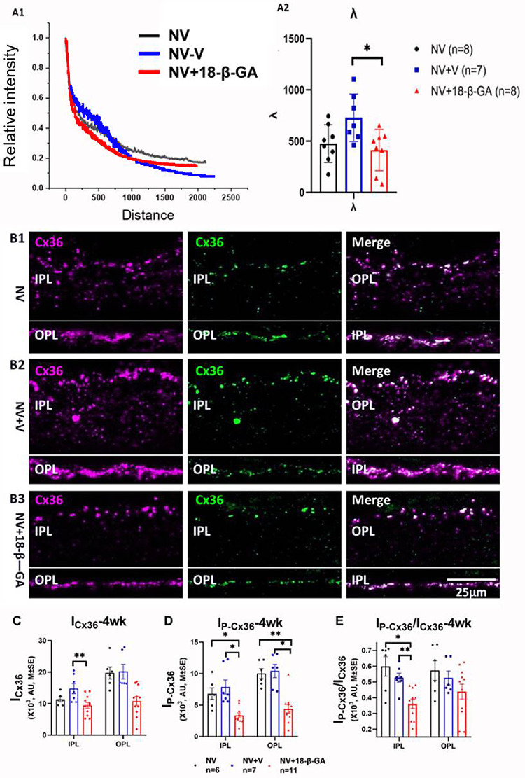
Results: FD per se (excluding effect of light-attenuation) increased HC-HC coupling in OPL, whereas HD did not affect it. HD for 2 days or 1 week had no significant effect on retinal content of Cx36 or P-Cx36. FD for 4 weeks decreased the total amounts of Cx36 and P-Cx36, and the P-Cx36/Cx36 ratio, in the IPL. Subconjunctival 18-β-GA induced myopia in NV eyes and increased the myopic shifts in FD eyes, while reducing the amounts of Cx36 and P-Cx36 in both the IPL and OPL.

Conclusions: These results suggest that cell-cell coupling via GJs containing Cx36 (particularly those in the IPL) plays a role in emmetropization and form deprivation myopia (FDM) in mammals. Although both FD and 18-β-GA induced myopia, they had opposite effects on HC-HC coupling. These findings suggest that HC-HC coupling in the OPL might not play a significant role in emmetropization and myopia development.


Conflict of interest statement

Disclosure: Z. Zhi, None; J. Xiang, None; Q. Fu, None; X. Pei, None; D. Zhou, None; Y. Cao, None; L. Xie, None; S. Zhang, None; S. Chen, None; J. Qu, None; X. Zhou, None

Figures

Figure 1.
Figure 1.
Experimental Design. HC, horizontal cell; FD, form deprivation; HD, hyperopic defocus; NV, normal visual environment; IF, immunofluorescence; GJs, gap junctions; EIR, eccentric infrared retinoscopy.
Figure 2.
Figure 2.
Procedure and analysis of the cut-loading technique. (A) Schematic diagram of cut-loading technique. (B) A representative example shows fluorescence of a flat-mounted guinea pig retina, following cut-loading with a solution of both neurobiotin (green, B1) and rhodamine dextran (magenta, B2). Rhodamine dextran labels only damaged cells along the cut, whereas neurobiotin diffuses through GJs between viable cells and can be seen in coupled cells far from the cut. (C) The neurobiotin diffuses to different extents in the Retinal Ganglion Cell Layer (RGL, C1), inner sublayer of Inner Nuclear Layer (inner INL, C2), and outer sublayer of INL (C3). In the RGL and inner INL, tracer coupling is limited mainly to a range of less than 300 µm from the cut. In the outer INL, a layer of cells is more extensively coupled (over 1000 µm from the cut). (D) The coupled cells in the outer INL (D1) are co-labeled for the horizontal cell marker, calbindin (D2, D3). (E) Tracer coupling of horizontal cells is revealed by diffusion of neurobiotin (E1); E2: Normalized relative fluorescence intensity of the horizontal cell layer as a function of distance from the cut.
Figure 3.
Figure 3.
The effects of FD, low illuminance, and HD on horizontal cell tracer coupling after 2 days’ treatment. (A) FD increased horizontal cell tracer coupling. (A1) An example of diffusion curves from animals raised in normal visual environment (NV) and the fellow (FD-F) and treated eyes (FD-T) of the FD animals. (A2) The space constants of each group (NV: n = 8, FD-F: n = 10, and FD-T: n = 10). (B) Illuminance of 40 lux decreased horizontal cell tracer coupling. (B1) Example of diffusion curves from 300 lux and 40 lux groups. (B2) Space constants of the two illuminance groups (300 lux: n = 6; 40 lux: n = 10). (C) HD did not affect horizontal cell tracer coupling. (C1) Example of diffusion curves from normal vision (NV, n = 8), 0 D (n = 6), and defocus (−4 D, −4 D-F: n = 8; −4 D-T: n = 11) groups. (C2) The space constants of the −4 D and 0 D groups. *P < 0.05.
Figure 4.
Figure 4.
Effects of HD on the fluorescence intensities of retinal Cx36 and P-Cx36 (M ± SE). (A) Fluorescence images of vertical sections of guinea pig retina in 0 D and −4 D groups for 2 days and 1 week. (A1) 0 D; (A2) −4 D; Left panel: HD for 2 days; Right panel: HD for 1 week. (B) Total fluorescence intensity of Cx36 in IPL and OPL after HD 2 days. (C) Total fluorescence intensity of P-Cx36 in IPL and OPL after HD 2 days. (D) Fluorescence intensity ratios (P-Cx36/Cx36) in IPL and OPL after HD 2 days. (E) Total fluorescence intensity of Cx36 in IPL and OPL after HD 1 week. (F) Total fluorescence intensity of P-Cx36 in IPL and OPL after HD 1 week. (G) Fluorescence intensity ratios (P-Cx36/Cx36) in IPL and OPL after HD 1 week. Wearing −4 D or 0 D lens for 2 days and 1 week did not significantly affect either the fluorescence intensity of Cx36 or P-Cx36, or the phosphorylation level of Cx36 (P-Cx36:Cx36), in either IPL or OPL. AU units represent relative fluorescence intensity.
Figure 5.
Figure 5.
FD for 2 days: The effects of FD and low illuminance on the fluorescence intensity of retinal Cx36 and P-Cx36 (M ± SE). (A) Fluorescence images of vertical sections of guinea pig retina of the FD-T (n = 7), NV-300 lux (n = 7), NV-40 lux (n = 7) groups. A1: Cx36; A2: P-Cx36; A3: Merged. (B) Total fluorescence intensity of Cx36 in IPL and OPL. (C) Total fluorescence intensity of P-Cx36 in IPL and OPL. (D) Fluorescence intensity ratios (P-Cx36/Cx36) in IPL and OPL. FD for 2 days did not significantly affect the total fluorescence intensities of Cx36 or P-Cx36 nor the phosphorylation levels of Cx36 (P-Cx36/Cx36), in either IPL or OPL, at either illuminance. AU units represent relative fluorescence intensity.
Figure 6.
Figure 6.
FD for 4 w ee ks reduced the levels of Cx36 and its phosphorylation in IPL and OPL (M ± SE). (A) Immunofluorescence of Cx36 and P-Cx36 in IPL and OPL. A1: NV (n = 9); A2: FD-T (n = 6). Magenta: Cx36; Green: P-Cx36; White: Merge. (B) FD decreased the total intensity of Cx36-IR plaques in IPL. (C) FD decreased the total intensity of P-Cx36-IR plaques in IPL. (D) FD decreased the phosphorylation level of Cx36 (IP-Cx36/ ICx36) in IPL. *P < 0.05, independent t-test. AU units represent relative fluorescence intensity.
Figure 7.
Figure 7.
Subconjunctival injection of 18-β-GA enhanced ocular elongation and myopia development, in normal vision (NV) and form-deprivation (FD) conditions (M ± SE). (A) Interocular differences of refraction (A1), vitreous chamber depth (VCD) (A2), and axial length (AL) (A3), in NV. (B) Interocular differences of refraction (B1), vitreous chamber depth (VCD) (B2) and axial length (AL) (B3), in FD. Injection of 18-β-GA induced myopia development in both NV (A1) and FD (B1). VCD and AL were consistently increased in NV (A2, A3), but not obviously in FD (B2, B3). * P < 0.05; ** P < 0.01; *** P < 0.001, 2-way repeated ANOVA, Bonferroni correction, post hoc test.
Figure 8.
Figure 8.
Effects of 18-β-GA on horizontal cell tracer coupling (at day 2), Cx36 protein content, and Cx36 phosphorylation (at week 4). Injection of 18-β-GA decreased horizontal cell tracer coupling (M ± SE) (A). (B) Immunofluorescence of Cx36 and P-Cx36 in the IPL and OPL of retinas treated by 18-β-GA-injection. (B1) NV (n = 13); (B2)NV + V (n = 7); (B3): NV + 18-β-GA (n = 11). Magenta: Cx36; Green: P-Cx36; White: Merged Cx36 + P-Cx36. Injection of 18-β-GA decreased the levels of Cx36 (C), P-Cx36 (D), and Cx36 phosphorylation (P-Cx36:Cx36) in IPL (E), in comparison to NV + V controls (M ± SE). * P < 0.05; ** P < 0.01; *** P < 0.001, 1-way ANOVA. AU units represent relative fluorescence intensity.

Similar articles

Cited by

References

    1. Holden BA, Fricke TR, Wilson DA, et al. .. Global prevalence of myopia and high myopia and temporal trends from 2000 through 2050. Ophthalmology. 2016; 123: 1036–1042. - PubMed
    1. Zhou X, Lu F, Xie R, et al. .. Recovery from axial myopia induced by a monocularly deprived facemask in adolescent (7-week-old) guinea pigs. Vision Res . 2007; 47: 1103–1111. - PubMed
    1. Zheng H, Tse DY, Tang X, To C, Lam TC.. The interactions between bright light and competing defocus during emmetropization in chicks. Invest Ophthalmol Vis Sci . 2018; 59: 2932–2943. - PubMed
    1. Xiong S, Sankaridurg P, Naduvilath T, et al. .. Time spent in outdoor activities in relation to myopia prevention and control: a meta-analysis and systematic review. Acta Ophthalmologica. 2017; 95: 551–566. - PMC - PubMed
    1. Wässle H. Parallel processing in the mammalian retina. Nat Rev Neurosci . 2004; 5: 747–757. - PubMed

Publication types

MeSH terms