. 2021 Jul 20;131(17):e146950.
doi: 10.1172/JCI146950. Online ahead of print.

Systemic inhibition of PTPN22 augments anticancer immunity

Affiliations

Systemic inhibition of PTPN22 augments anticancer immunity

Won Jin Ho et al. J Clin Invest. .

Abstract

Both epidemiologic and cellular studies in the context of autoimmune diseases have established that protein tyrosine phosphatase non-receptor type 22 (PTPN22) is a key regulator of T cell receptor (TCR) signaling. However, its mechanism of action in tumors and its translatability as a target for cancer immunotherapy have not been established. Here we show that a germline variant of PTPN22, rs2476601, portended a lower likelihood of cancer in patients. PTPN22 expression was also associated with markers of immune regulation in multiple cancer types. In mice, lack of PTPN22 augmented antitumor activity with greater infiltration and activation of macrophages, natural killer (NK) cells, and T cells. Notably, we generated a novel small molecule inhibitor of PTPN22, named L-1, that phenocopied the antitumor effects seen in genotypic PTPN22 knockout. PTPN22 inhibition promoted activation of CD8+ T cells and macrophage subpopulations toward MHC-II expressing M1-like phenotypes, both of which were necessary for successful antitumor efficacy. Increased PD1-PDL1 axis in the setting of PTPN22 inhibition could be further leveraged with PD1 inhibition to augment antitumor effects. Similarly, cancer patients with the rs2476601 variant responded significantly better to checkpoint inhibitor immunotherapy. Our findings suggest that PTPN22 is a druggable systemic target for cancer immunotherapy.

Keywords: Cancer immunotherapy; Drug therapy; Immunology; Oncology.


Conflict of interest statement

Conflict of interest: WH, SC, JL, ZYZ, BHP, and EMJ are co-inventors on invention disclosures related to targeting PTPN22 to treat cancers (WO2021007491A1), developing novel compounds against PTPN22, and/or utilizing rs2476601 as a predictive biomarker for cancer immunotherapy (disclosed). WH is a co-inventor on patents with potential for receiving royalties from Rodeo Therapeutics. MY reports receiving research grants from Incyte, Bristol-Myers Squibb, and Exelixis, and is a consultant for AstraZeneca, Eisai, Exelixis, and Genentech. EJF is a consultant for Champions Oncology. BHP had ownership interest and was a paid member of the scientific advisory board of Loxo Oncology and was a paid consultant for Foundation Medicine, Inc, Lilly, Casdin Capital, and Roche. He is currently a paid scientific advisory board member of Celcuity and a paid consultant for Jackson Laboratories and Pathovax. Under separate licensing agreements between Horizon Discovery, LTD and The Johns Hopkins University, BHP is entitled to a share of royalties received by the University on sales of products. The terms of this arrangement are being managed by the Johns Hopkins University in accordance with its conflict of interest policies. ZYZ is a co-founder and serves on the scientific advisory board of Tyligand Bioscience. EMJ reports receiving a commercial research grant from Bristol-Myers Squibb

Figures

Figure 1
Figure 1. PTPN22 is associated with a negative regulatory role in the immune response against cancer.
(A) A volcano plot showing the results from the analysis using phenome-wide association studies (PheWAS) from the Vanderbilt BioVU database (n = 72,083). Each dot represents an association between Ptpn22 rs2476601 and a disease diagnosis. The horizontal dashed line indicates an FDR-adjusted P value of 0.05. (B) Correlations between Ptpn22 expression and immune cell types deconvolved by CIBERSORT across 11 cancer types from The Cancer Genome Atlas (TCGA) are shown as a heatmap. (C) Correlation between Ptpn22 and immune regulatory markers across 11 cancer types from TCGA are shown as a heatmap. BLCA, bladder cancer; BRCA, breast cancer; COAD, colon adenocarcinoma; GBM, glioblastoma multiforme; HNSC, head and neck squamous cell cancer; LIHC, hepatocellular carcinoma; LUAD, lung adenocarcinoma; LUSC, lung squamous cell carcinoma; PAAD, pancreatic ductal adenocarcinoma; PRAD, prostate adenocarcinoma; SKCM, skin melanoma.
Figure 2
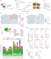
Figure 2. PTPN22 KO confers protection against MC38 tumor growth in association with enhanced immune infiltration.
(A) Schematic of the mouse tumor model used. Cells (2.5 × 10⁵) were injected subcutaneously in the right hind limb for tumor growth measurements 2 to 3 times a week through 21 days after injection. (B) MC38 tumor growth was compared between WT (blue circles) and PTPN22-KO (red squares) mice (n = 10–11 per arm). ***P < 0.001 by nonlinear regression. Pictures on the adjacent panel show gross morphology of the 2 representative tumors from each arm. (C) Weights of the MC38 tumors on the day of the harvest (day 21) from WT and PTPN22-KO mice (n = 15–16). (D and E) Immunohistochemical analysis of the tumors comparing CD4+ and CD8+ cells and Foxp3+ cells infiltrating the tumors (scale bars: 100 μm). Positive staining was quantified using HALO software (n = 11–13 per arm). Foxp3+ density is represented as a proportion of T cell density (sum of CD4+ and CD8+ densities). *P < 0.05, ***P < 0.005 by 2-tailed, unpaired t test. (F and G) Immune subsets and T cell subsets are represented as percentage of live cells (WT vs. PTPN22-KO, mean ± SEM, n = 8). *P < 0.05, **P < 0.01, ***P < 0.005. UMAP plot from CyTOF analysis of the immune profile is shown. Detailed annotations of cell types are shown in Supplemental Figure 2A. (H) Conventional flow cytometry was performed to validate CyTOF findings showing increased CD8+ T cells and CD4+ T cells within the MC38 tumors in PTPN22-KO mice compared with WT (n = 4). *P < 0.05, ***P < 0.005 by 1-tailed, unpaired t test. Ckpt, checkpoint markers; DC, dendritic cells; G-MDSC/TAN, granulocytic myeloid-derived suppressor cells/tumor associated neutrophils; Gz, granzyme; M-MDSC/MC, monocytic myeloid-derived suppressor cells/myeloid cells; MC, monocytes; NK, natural killer cells; NKT, natural killer T cells; TAM, tumor-associated macrophages; Tc, cytotoxic T cells; Th, helper T cells; Treg, regulatory T cells.
Figure 3
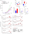
Figure 3. Treatment with a small molecule inhibitor of PTPN22, L-1, phenocopies PTPN22 KO.
(A) Schematic of the L-1 treatment of MC38 and CT26 tumor model. Starting on day 3 of injection, L-1 is administered intraperitoneally twice daily for 5 consecutive days per week for 2 weeks and once daily for 5 consecutive days per week for 1 week. The structure of L-1 is illustrated. (B and C) Tumor growth of MC38 in C57BL/6J and CT26 in BALB/cJ was compared between vehicle (blue circles) and L-1 (red squares) treatment groups. Mean ± SEM (n = 9–10 per arm). ***P < 0.001 by nonlinear regression. (D) Immunohistochemical analysis of the MC38 tumors shows CD4+ and CD8+ cells infiltrating the tumors (scale bars: 100 μm). Positive staining was quantified using HALO software, and the results are shown as mean ± SEM (n = 10–13 per arm). *P < 0.05 by 1-tailed, unpaired t test. (E) To assess potential off-target effects of L-1, starting on day 3 of injection, WT or PTPN22-KO mice were given either vehicle (VEH) or L-1 intraperitoneally. Tumor growth curves for WT VEH (blue inverted triangles), WT L-1 (red triangles), PTPN22-KO VEH (green squares), and PTPN22-KO L-1 (purple circles). Mean ± SEM (n = 4–5 per arm). **P < 0.01; ***P < 0.005 by nonlinear regression; not significant between PTPN22-KO VEH and PTPN22-KO L-1.
Figure 4
Figure 4. Antitumor effects of PTPN22 inhibition are mediated by CD8+ T cells.
(A) Tumor growth was compared across 6 groups: WT or PTPN22-KO mice treated with isotype, anti-CD4, or anti-CD8 antibodies (n = 4–5). Representative of 2 independent runs. *P < 0.05, ***P < 0.005 by nonlinear regression. (B and C) TCR repertoires for T cells infiltrating MC38 tumors were compared for WT vs. PTPN22-KO and VEH vs. L-1 treatments by TCRseq based on Shannon’s entropy and sample clonality (n = 4–5). *P < 0.05. (D) Tumor resistance experiment with EG7 tumors: 2.5 × 10⁵ cells were injected subcutaneously in the right hind limb, and tumor persistence was assessed on day 35. The frequency of tumors rejected in WT and PTPN22-KO mice is displayed (n = 20). (E) SIINFEKL tetramer+ CD8+ T cells in the tumor-draining lymph nodes from EG7 tumor–bearing mice were compared by flow cytometry using 1-way ANOVA followed by pairwise Tukey’s test (n = 9). *P < 0.05. (F) SIINFEKL tetramer+ CD8+ T cells from the spleens of vaccinated mice (n = 5). *P < 0.05, ***P < 0.005 by 1-way ANOVA followed by pairwise Tukey’s test. (G) Phosphorylation intensities (mean metal intensities) for each of the indicated phospho-site, stratified by the subtype of CD8+ T cells, comparing MC38 tumor–infiltrating CD8+ T cells from WT and PTPN22-KO mice (n = 5). In the box-and-whisker plots in B, C, and G, the bottom and top hinges of the boxes mark the 25th and 75th percentiles, respectively, and the lines within the boxes are medians. Whiskers represent 1.5 times the interquartile range extending from the hinges. Results of linear mixed modeling for differential analyses of the phosphorylation levels are shown as FDR-adjusted P values: *P < 0.1, ***P < 0.005. Detailed annotations of CD8+ T cell clusters are shown in Supplemental Figure 7. CM, central memory subtype; EM, effector memory subtype; Eff, effector subtype; EX, exhausted subtype (positive expression of checkpoint markers).
Figure 5
Figure 5. Remodeling of F4/80+ TAM compartment mediates the antitumor efficacy of L-1.
(A) Two independent replicate runs testing the effects of VEH vs. L-1 with or without anti-F4/80 on MC38 tumor growth are shown (n = 3–5 for run 1 [left] and n = 5 for run 2 [right]). *P < 0.05, ***P < 0.005 by nonlinear regression. (BE) CyTOF analysis of the F4/80+ populations in the MC38 model. (B) The extent of anti-F4/80 depletion observed within the tumors. ***P < 0.005 only for the anti-F4/80 effect based on 2-way ANOVA. (C) Correlations among phenotypic markers relevant to TAMs within the TAM data subset. (D) Hierarchically clustered expression heatmap of annotated TAM clusters. (E) The proportion of TAM and T cell subpopulations within the tumors across the 4 groups (n = 5–10). *P < 0.05, **P < 0.01, ***P < 0.005 by 1-way ANOVA.
Figure 6
Figure 6. Targeting PTPN22 synergizes anti–PD-1 therapy.
(A) Tumor growth experiment testing the effects of a single 200-μg dose (day 8) of anti–PD-1 therapy against MC38 tumors in WT and PTPN22-KO mice. Growth curves are shown for 4 groups: WT or PTPN22-KO mice treated with isotype antibody (WT ISO or PTPN22 KO ISO) or anti–PD-1 antibody (WT anti–PD-1 or PTPN22 anti–PD-1) (n = 5). ***P < 0.005 by nonlinear regression. (B and C) Combination of PTPN22 inhibitor L-1 with or without anti–PD-1 therapy (n = 9–10) in MC38 (B) or CT26 (C) models. *P < 0.05, **P < 0.01, ***P < 0.005 by nonlinear regression. (D) Response to checkpoint immunotherapy in patients with rs2476601 or WT Ptpn22 from the BioVU database. “Combination Therapy” (gray dots) refers to a combination of the indicated immunotherapy with a nonimmunotherapeutic drug. Inset: Kaplan-Meier curves comparing progression-free survival.

References

    1. Rosenberg SA. Decade in review-cancer immunotherapy: entering the mainstream of cancer treatment. Nat Rev Clin Oncol. 2014;11(11):630–632. doi: 10.1038/nrclinonc.2014.174. - DOI - PMC - PubMed
    1. Fesnak AD, et al. Engineered T cells: the promise and challenges of cancer immunotherapy. Nat Rev Cancer. 2016;16(9):566–581. doi: 10.1038/nrc.2016.97. - DOI - PMC - PubMed
    1. Pardoll DM. The blockade of immune checkpoints in cancer immunotherapy. Nat Rev Cancer. 2012;12(4):252–264. doi: 10.1038/nrc3239. - DOI - PMC - PubMed
    1. Ribas A, Wolchok JD. Cancer immunotherapy using checkpoint blockade. Science. 2018;359(6382):1350–1355. doi: 10.1126/science.aar4060. - DOI - PMC - PubMed
    1. Bottini N, Peterson EJ. Tyrosine phosphatase PTPN22: multifunctional regulator of immune signaling, development, and disease. Annu Rev Immunol. 2014;32:83–119. doi: 10.1146/annurev-immunol-032713-120249. - DOI - PMC - PubMed