. 2021 Jul 20;17(7):e1009738.
doi: 10.1371/journal.ppat.1009738. eCollection 2021 Jul.

Improved delivery of broadly neutralizing antibodies by nanocapsules suppresses SHIV infection in the CNS of infant rhesus macaques

Affiliations

Improved delivery of broadly neutralizing antibodies by nanocapsules suppresses SHIV infection in the CNS of infant rhesus macaques

Jing Wen et al. PLoS Pathog. .

Abstract

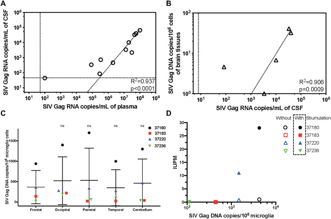
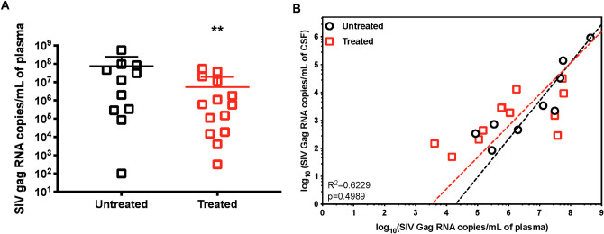
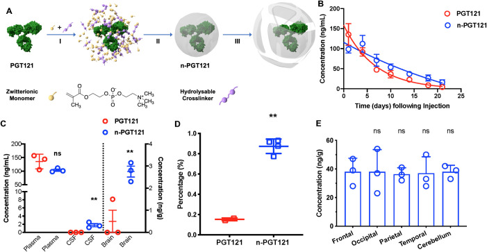
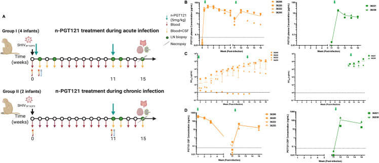
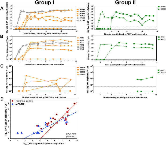
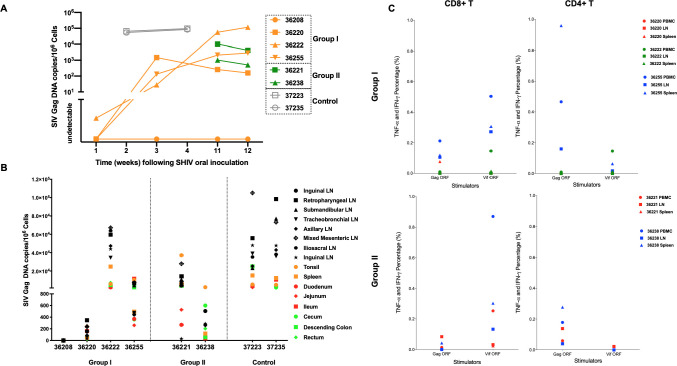
Broadly neutralizing antibodies (bNAbs) directed to HIV-1 have shown promise at suppressing viremia in animal models. However, the use of bNAbs for the central nervous system (CNS) infection is confounded by poor penetration of the blood brain barrier (BBB). Typically, antibody concentrations in the CNS are extremely low; with levels in cerebrospinal fluid (CSF) only 0.1% of blood concentrations. Using a novel nanotechnology platform, which we term nanocapsules, we show effective transportation of the human bNAb PGT121 across the BBB in infant rhesus macaques upon systemic administration up to 1.6% of plasma concentration. We demonstrate that a single dose of PGT121 encased in nanocapsules when delivered at 48h post-infection delays early acute infection with SHIVSF162P3 in infants, with one of four animals demonstrating viral clearance. Importantly, the nanocapsule delivery of PGT121 improves suppression of SHIV infection in the CNS relative to controls.


Conflict of interest statement

I have read the journal’s policy and the authors of this manuscript have the following competing interests: I.S.Y.C. has a financial interest in CSL Behring and Calimmune Inc. J.W. has a financial interest in Vivibaba and The Regents have licensed intellectual property invented by J.W. to Vivibaba. Y.L. has a financial interest in Vivibaba and The Regents have licensed intellectual property invented by Y.L. to Vivibaba. No funding was provided by these companies to support this work.

Figures

Fig 1
Fig 1. Infection occurs in the central nervous system (CNS) of infant rhesus macaques.
11 one-month-old infant rhesus macaques were exposed to a single high dose SHIVSF162P3 challenge by atraumatic oral inoculation. A) Correlation between SHIVSF162P3 Gag RNA copies in plasma and CSF in infected infant rhesus macaques. The animals showing positive RNA copies in both plasma and CSF are included in the figure. Dotted lines indicate limits of detection. Each dot represents one animal, and all data are fitted by nonlinear log-log line regression. R2 and p value indicate the fitness of Pearson correlation. Dotted lines indicate limits of detection. B) Correlation between SHIVSF162P3 Gag RNA copies in CSF and vDNA copies in brain tissue. Animals showing positive RNA copies in both CSF and vDNA are included in the figure. Each dot represents one animal, and all data are fitted by nonlinear log-log line regression. R2 and p value indicate the fitness of Pearson correlation. Dotted lines indicate limits of detection. C) Comparable viral DNA copies in microglia cells in five brain locations. The mean of vDNA copies in microglia from five brain locations are compatible. Data are shown as means ± s.d. of four animals. Statistical significance, compared with the vDNA in microglia from Frontal, was determined by two-tailed unpaired t test. ns: not significant. D) Inducible virus is correlated with vDNA in microglia. The inducible virus was detected viaTZA assay. IUPM, infectious units per million microglia cells. Primary microglia were stimulated with 20 ng/mL M-CSF and 25 ng/mL IL-10 for 5 days. Both Without and With Stimulation fit Pearson correlation. Without Stimulation: R2 = 0.771, p = 0.1219; With Stimulation: R2 = 0.9854, p = 0.0073.
Fig 2
Fig 2. The bNAb-based treatment impacts infection in blood but not in the CNS in infant rhesus macaques.
14 one-month-old infant rhesus macaques were exposed to a single high dose SHIVSF162P3 challenge by atraumatic oral inoculation and treated with bNAbs or cART+bNAbs with various doses and regimens at 48h post-infection (details in S1 Table). A) Viral RNA copies in plasma of infant rhesus macaques without treatment (Untreated) and treated with bNAbs or bNAbs+cART (Treated) at 48 h. Statistical significance was calculated to untreated group by two-tailed unpaired t test. **: P values < 0.0021. B) Comparison of correlations between viral RNA copies in plasma and CSF of Untreated and Treatment animals. Each dot represents one animal, and all data are fitted by nonlinear log-log line regression. Significance between the correlations of two groups is calculated by multiple regression model. There is no statistically significant difference between slopes (p-value = 0.4989).
Fig 3
Fig 3. Nanocapsules improve bNAb penetration in CNS.
A) Scheme of the synthesis and release of PGT121 nanocapsules (n-PGT121) by (I) enriching zwitterionic monomer (MPC) and hydrolysable crosslinkers (PLA-PEG-PLA) around a PGT121 molecule, (II) in-situ polymerization of the monomer and crosslinkers forming a thin shell of polymer around PGT121, and (III) releasing PGT121 when polymer shells are degraded under physiological condition by hydrolysis of the crosslinkers. B) Kinetics of native PGT121 and n-PGT121 in mice. C57BL/6 mice were randomly separated into two groups and injected with 5 mg/kg of PGT121 (n = 6) and n-PGT121 (n = 6) via retro-orbital injection. The release of PGT121 was assayed by ELISA using ST09 and anti-human IgG. C) Nanocapsules improved CNS delivery (CSF and brain tissue) of PGT121 in mice. C57BL/6 mice were randomly separated into two groups and injected with 5 mg/kg of PGT121 (n = 6) and n-PGT121 (n = 6) via retro-orbital injection. Body fluids and tissues were collected 3 days after injection. The concentrations of native and released PGT121 in plasma, CSF, and perfused brain homogenates were measured by ELISA. Data are shown as means ± s.d. of three animals. Statistical significance was calculated to the PGT121 group by two-tailed unpaired t test. ns: not significant; **: P values < 0.0021. D) SHIV infected infant rhesus macaques were injected with 5 mg/kg PGT121. The percentage of PGT121 concentration in CSF of that in plasma of infant rhesus macaques. The percentages of PGT121 concentration in CSF and that in plasma from infant rhesus macaques treated with native PGT121 (n = 2) and n-PGT121 (n = 4) on Day 7 after injection. The concentrations of native and released PGT121 in CSF were measured by ELISA. Data are shown as means ± s.d. of each group. Statistical significance was calculated to PGT121 group by two-tailed unpaired t test. **: P values< 0.0021. E) Nanocapsules show a uniform distribution in different brain locations of rhesus macaques. Five brain tissues from n-PGT121 treated SHIV infected infant rhesus macaques were weighted, washed, and homogenized. Supernatant was collected after centrifuging the homogenate. The concentration of released PGT121 was detected by ELISA. Statistical significance was calculated to Frontal by two-tailed unpaired t test. ns: not significant.
Fig 4
Fig 4. Nanocapsules maintain bNAb circulation and improve penetration in CNS in SHIV-infected infant rhesus macaques.
A) Experimental design of the n-PGT121 treatment in SHIV infected infant rhesus macaques at 48 h during acute infection (Group I) and Week 11 (WK11) during chronic infection (Group II). One-month-old infant rhesus macaques negative for Mamu-B*08 and Mamu-B*17 alleles were randomly assigned to groups. All animals were exposed to a single high dose SHIVSF162P3 challenge by atraumatic oral inoculation. Black arrows indicate SHIV infection. Green arrows indicate n-PGT121 treatments. Dark red arrows indicate blood collection. Dark yellow arrows indicate CSF collection. Green circles indicate LN biopsies. Scalpels indicate time of necropsy. B) The released PGT121 concentration from n-PGT121 in plasma in acute infection group (Group I, n = 4) and chronic infection group (Group II, n = 2). WK0 and WK11 samples were collected before n-PGT121 infusion. Data are shown as means ± s.d. of each group. Green arrows indicate n-PGT121 treatments. C) Seroconversion was determined by measuring the concentration (EC50) of envelope antibodies in the plasma of the acute infection group (Group I, n = 4) and chronic infection group (Group II, n = 2). EC50 was assayed by an SHIVSF162 gp140-specific ELISA. Data are shown as individual data for each animal and means ± s.d. of each group. Pre-treatment plasma (day 0) was used as a negative control for the assay. WK11 samples for this assay were collected before n-PGT121 infusion. Green arrows indicate n-PGT121 treatments. D) The released PGT121 concentration from n-PGT121 in CSF in acute infection group (Group I, n = 4) and chronic infection group (Group II, n = 2). WK0 and WK11 samples were collected before n-PGT121 infusion. Data are shown as means ± s.d. of each group. Green arrows indicate n-PGT121 treatments.
Fig 5
Fig 5. Nanocapsules reduce viremia in both plasma and CNS of SHIV-infected infant rhesus macaques.
A) Plasma viral loads (viral RNA copies), B) the PBMC-associated viral loads (vDNA), and C) the CSF viral loads (viral RNA copies) were measured longitudinally in n-PGT121 treated animals in the acute infection group (Group I, n = 4) and chronic infection group (Group II, n = 2). Key to individual animals is shown. D) The comparison between Historical Control and n-PGT121 animals on correlation between viral RNA copies in plasma and CSF. Each dot represents one animal, and all data are fitted by nonlinear log-log line regression. The significance between the correlations of two groups is calculated with a multiple regression model. A statistically significant difference between slopes is observed in the multiple regression model (p-value = 0.0020). Both untreated and treated animals with native bNAbs or bNAbs+cART are combined and plotted as Historical Control (Untreated and Treated in Fig 2B).
Fig 6
Fig 6. Impact of bNAb nanocapsules on tissue-associated viremia is consistent with plasma viremia.
A) Cell-associated viral loads (vDNA) were measured in lymph node biopsies longitudinally in untreated control, Group I, and Group II animals. B) SHIV DNA was quantified by ultrasensitive nested qPCR in all tissue samples from the untreated control, Group I, and Group II animals at necropsy. Tissues are color coded in the figure. Black symbols represent lymph nodes from different locations. Red symbols represent tissues in the small intestine. Green symbols represent tissues in the large intestine. Orange symbols represent the other lymphatic tissues, including tonsil and spleen. C) CD8+ and CD4+ T cell responses to SIVmac239 Gag ORF 15-mer peptide pool, and SIVmac239 Vif ORF 15-mer peptide pool were measured by flow cytometric intracellular cytokine staining (ICS). Cells without stimulator were used as negative controls. Different tissue types (PBMC, LN from mesentery, spleen) are indicated with individual symbols as shown and the animal number is indicated next to the symbol.

Similar articles

Cited by

References

    1. Grant I, Atkinson JH, Hesselink JR, Kennedy CJ, Richman DD, Spector SA, et al.. Evidence for early central nervous system involvement in the acquired immunodeficiency syndrome (AIDS) and other human immunodeficiency virus (HIV) infections. Studies with neuropsychologic testing and magnetic resonance imaging. Ann Intern Med. 1987;107(6):828–36. doi: 10.7326/0003-4819-107-6-828 - DOI - PubMed
    1. Kaul M, Garden GA, Lipton SA. Pathways to neuronal injury and apoptosis in HIV-associated dementia. Nature. 2001;410(6831):988–94. doi: 10.1038/35073667 - DOI - PubMed
    1. Koyanagi Y, Miles S, Mitsuyasu RT, Merrill JE, Vinters HV, Chen IS. Dual infection of the central nervous system by AIDS viruses with distinct cellular tropisms. Science. 1987;236(4803):819–22. doi: 10.1126/science.3646751 - DOI - PubMed
    1. Major EO, Rausch D, Marra C, Clifford D. HIV-associated dementia. Science. 2000;288(5465):440–2. doi: 10.1126/science.288.5465.439d - DOI - PubMed
    1. Epstein LG, Kuiken C, Blumberg BM, Hartman S, Sharer LR, Clement M, et al.. HIV-1 V3 domain variation in brain and spleen of children with AIDS: tissue-specific evolution within host-determined quasispecies. Virology. 1991;180(2):583–90. doi: 10.1016/0042-6822(91)90072-j - DOI - PubMed

Publication types