

NLRP3 inhibition attenuates early brain injury and delayed cerebral vasospasm after subarachnoid hemorrhage
- PMID: 34284798
- PMCID: PMC8293512
- DOI: 10.1186/s12974-021-02207-x
NLRP3 inhibition attenuates early brain injury and delayed cerebral vasospasm after subarachnoid hemorrhage
Abstract
Background: The NLRP3 inflammasome is a critical mediator of several vascular diseases through positive regulation of proinflammatory pathways. In this study, we defined the role of NLRP3 in both the acute and delayed phases following subarachnoid hemorrhage (SAH). SAH is associated with devastating early brain injury (EBI) in the acute phase, and those that survive remain at risk for developing delayed cerebral ischemia (DCI) due to cerebral vasospasm. Current therapies are not effective in preventing the morbidity and mortality associated with EBI and DCI. NLRP3 activation is known to drive IL-1β production and stimulate microglia reactivity, both hallmarks of SAH pathology; thus, we hypothesized that inhibition of NLRP3 could alleviate SAH-induced vascular dysfunction and functional deficits.
Methods: We studied NLRP3 in an anterior circulation autologous blood injection model of SAH in mice. Mice were randomized to either sham surgery + vehicle, SAH + vehicle, or SAH + MCC950 (a selective NLRP3 inhibitor). The acute phase was studied at 1 day post-SAH and delayed phase at 5 days post-SAH.
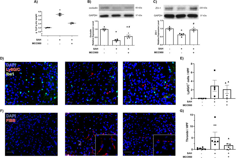
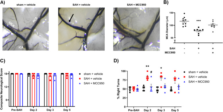
Results: NLRP3 inhibition improved outcomes at both 1 and 5 days post-SAH. In the acute (1 day post-SAH) phase, NLRP3 inhibition attenuated cerebral edema, tight junction disruption, microthrombosis, and microglial reactive morphology shift. Further, we observed a decrease in apoptosis of neurons in mice treated with MCC950. NLRP3 inhibition also prevented middle cerebral artery vasospasm in the delayed (5 days post-SAH) phase and blunted SAH-induced sensorimotor deficits.
Conclusions: We demonstrate a novel association between NLRP3-mediated neuroinflammation and cerebrovascular dysfunction in both the early and delayed phases after SAH. MCC950 and other NLRP3 inhibitors could be promising tools in the development of therapeutics for EBI and DCI.
Keywords: Inflammation; Microglia; NLRP3 inflammasome; Stroke; Subarachnoid hemorrhage.
© 2021. The Author(s).
Conflict of interest statement
The authors declare that they have no competing interests.
Figures


Similar articles
-
Hydrogen-Rich Saline Attenuated Subarachnoid Hemorrhage-Induced Early Brain Injury in Rats by Suppressing Inflammatory Response: Possible Involvement of NF-κB Pathway and NLRP3 Inflammasome.Mol Neurobiol. 2016 Jul;53(5):3462-3476. doi: 10.1007/s12035-015-9242-y. Epub 2015 Jun 20. Mol Neurobiol. 2016. PMID: 26091790
-
MCC950 attenuated early brain injury by suppressing NLRP3 inflammasome after experimental SAH in rats.Brain Res Bull. 2019 Mar;146:320-326. doi: 10.1016/j.brainresbull.2019.01.027. Epub 2019 Feb 1. Brain Res Bull. 2019. PMID: 30716395
-
Effects of NLRP3 inflammasome blockade on postresuscitation cerebral function in a rat model of cardiopulmonary resuscitation.Biomed Pharmacother. 2021 Nov;143:112093. doi: 10.1016/j.biopha.2021.112093. Epub 2021 Aug 30. Biomed Pharmacother. 2021. PMID: 34474352
-
Cerebrovascular pathophysiology of delayed cerebral ischemia after aneurysmal subarachnoid hemorrhage.Histol Histopathol. 2021 Feb;36(2):143-158. doi: 10.14670/HH-18-253. Epub 2020 Sep 30. Histol Histopathol. 2021. PMID: 32996580 Review.
-
Delayed Cerebral Ischemia After Subarachnoid Hemorrhage: Experimental-Clinical Disconnect and the Unmet Need.Neurocrit Care. 2020 Feb;32(1):238-251. doi: 10.1007/s12028-018-0650-5. Neurocrit Care. 2020. PMID: 30671784 Free PMC article.
Cited by
-
Emerging trends and hot spots of NLRP3 inflammasome in neurological diseases: A bibliometric analysis.Front Pharmacol. 2022 Sep 7;13:952211. doi: 10.3389/fphar.2022.952211. eCollection 2022. Front Pharmacol. 2022. PMID: 36160384 Free PMC article.
-
SIRT1 Promotes M2 Microglia Polarization via Reducing ROS-Mediated NLRP3 Inflammasome Signaling After Subarachnoid Hemorrhage.Front Immunol. 2021 Nov 24;12:770744. doi: 10.3389/fimmu.2021.770744. eCollection 2021. Front Immunol. 2021. PMID: 34899720 Free PMC article.
-
Pharmacological intervention of curcumin via the NLRP3 inflammasome in ischemic stroke.Front Pharmacol. 2023 Oct 17;14:1249644. doi: 10.3389/fphar.2023.1249644. eCollection 2023. Front Pharmacol. 2023. PMID: 37915409 Free PMC article. Review.
-
Inhibition of P2X4R attenuates white matter injury in mice after intracerebral hemorrhage by regulating microglial phenotypes.J Neuroinflammation. 2021 Aug 23;18(1):184. doi: 10.1186/s12974-021-02239-3. J Neuroinflammation. 2021. PMID: 34425835 Free PMC article.
-
Protective effects of histone deacetylase 6 specific inhibitor tubastatin A on subarachnoid hemorrhage in rats and the underlying mechanisms.Zhong Nan Da Xue Xue Bao Yi Xue Ban. 2023 Feb 28;48(2):172-181. doi: 10.11817/j.issn.1672-7347.2023.220167. Zhong Nan Da Xue Xue Bao Yi Xue Ban. 2023. PMID: 36999463 Free PMC article. Chinese, English.
References
MeSH terms
Substances
Grants and funding
LinkOut - more resources
Full Text Sources
