. 2021 Jul 20;14(1):38.
doi: 10.1186/s13039-021-00555-0.

Clinical significance and mechanisms associated with segmental UPD

Affiliations

Clinical significance and mechanisms associated with segmental UPD

Peter R Papenhausen et al. Mol Cytogenet. .

Abstract

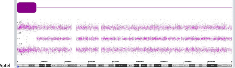
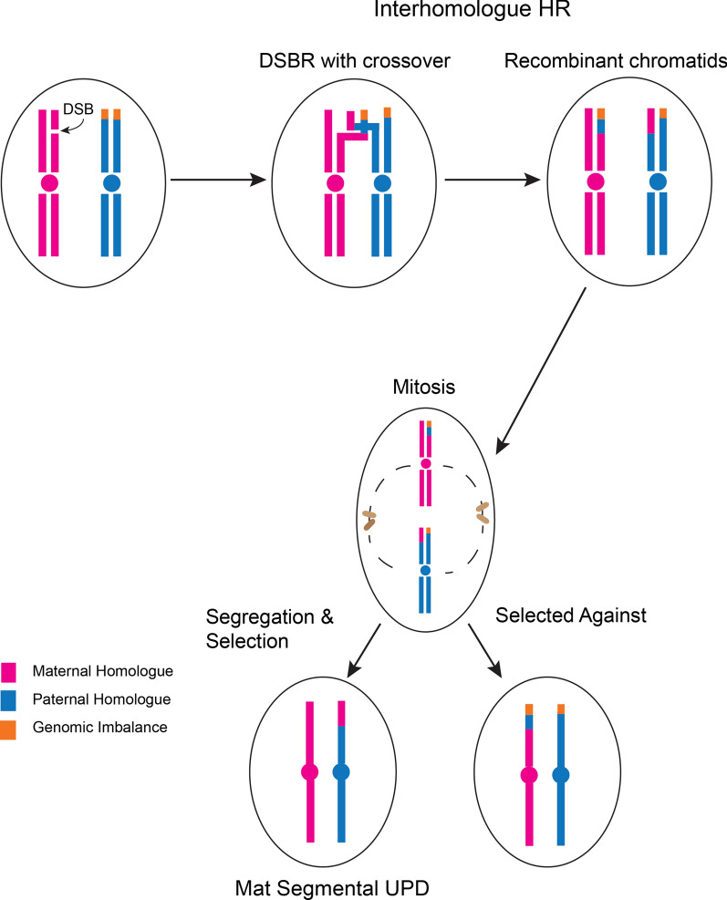
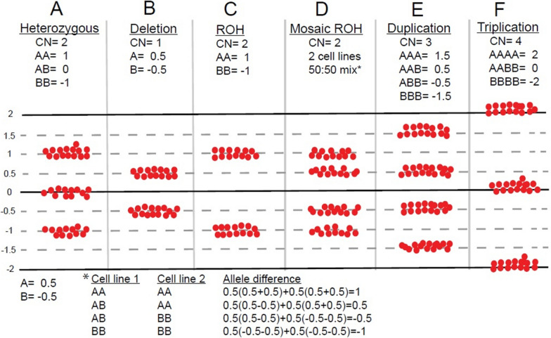
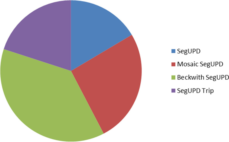
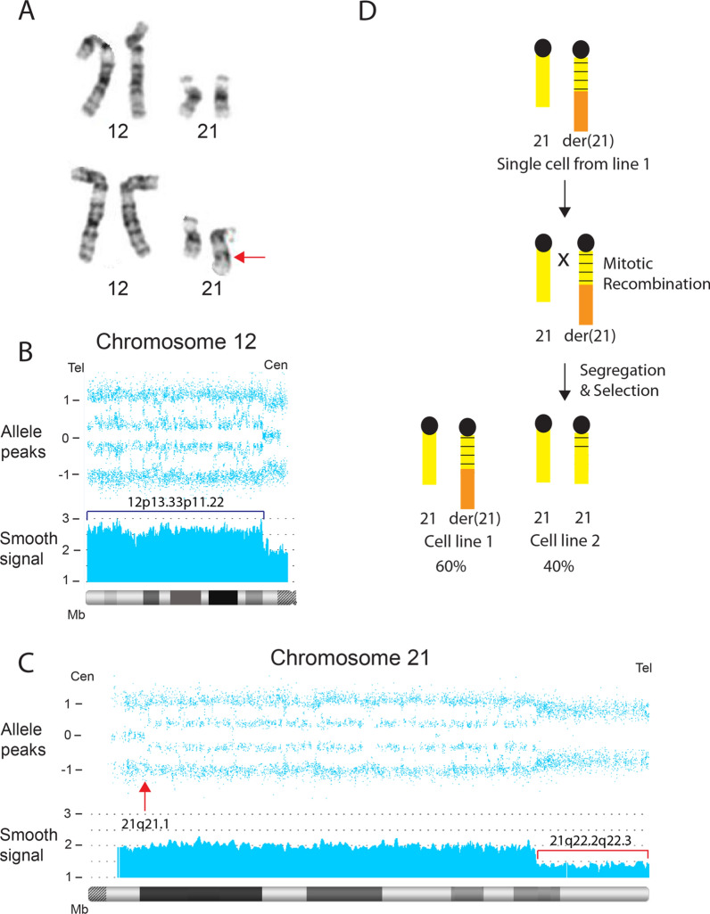
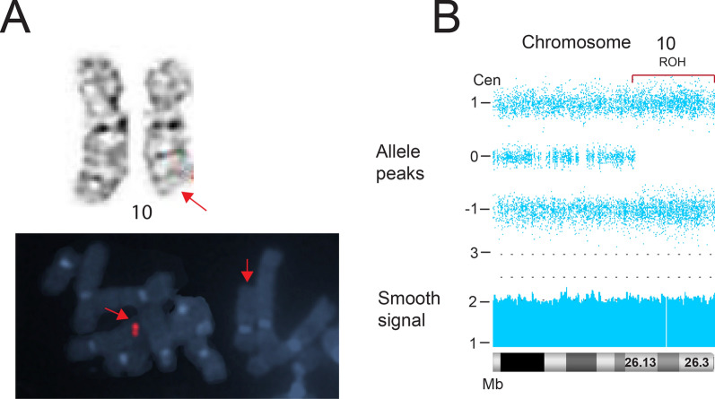
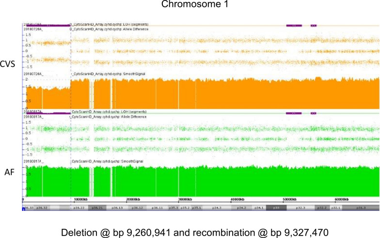
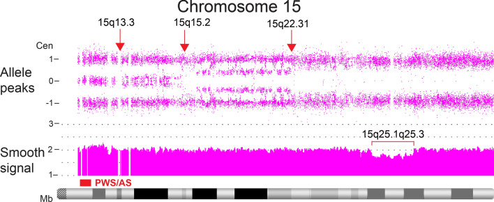
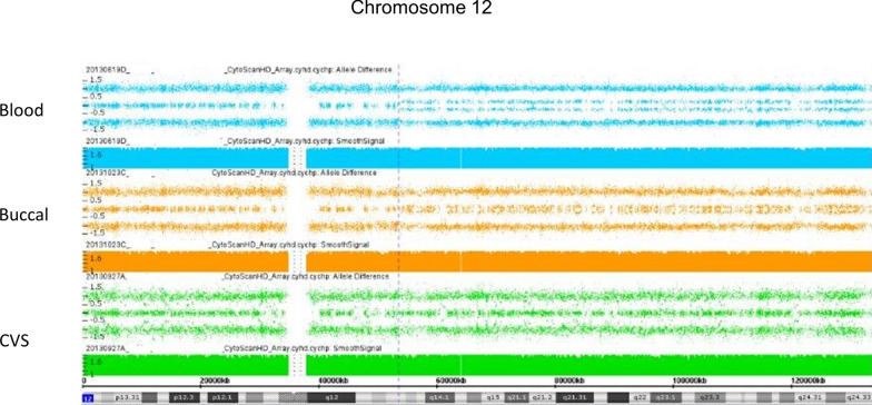
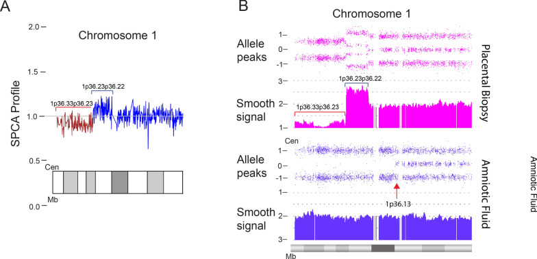
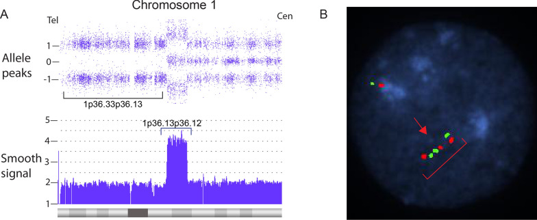
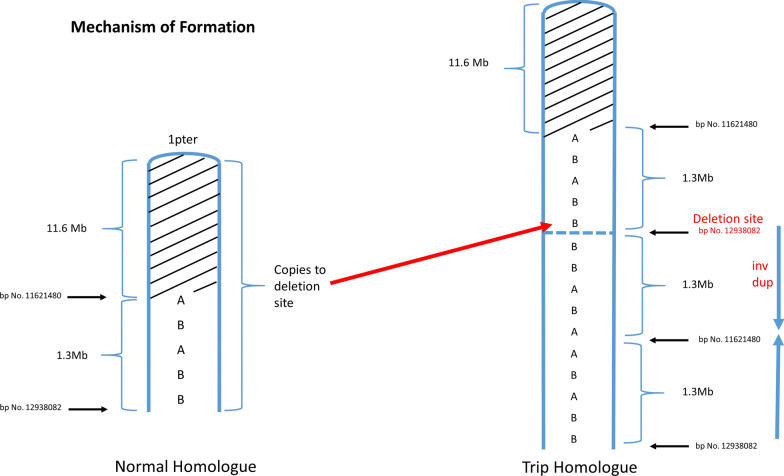
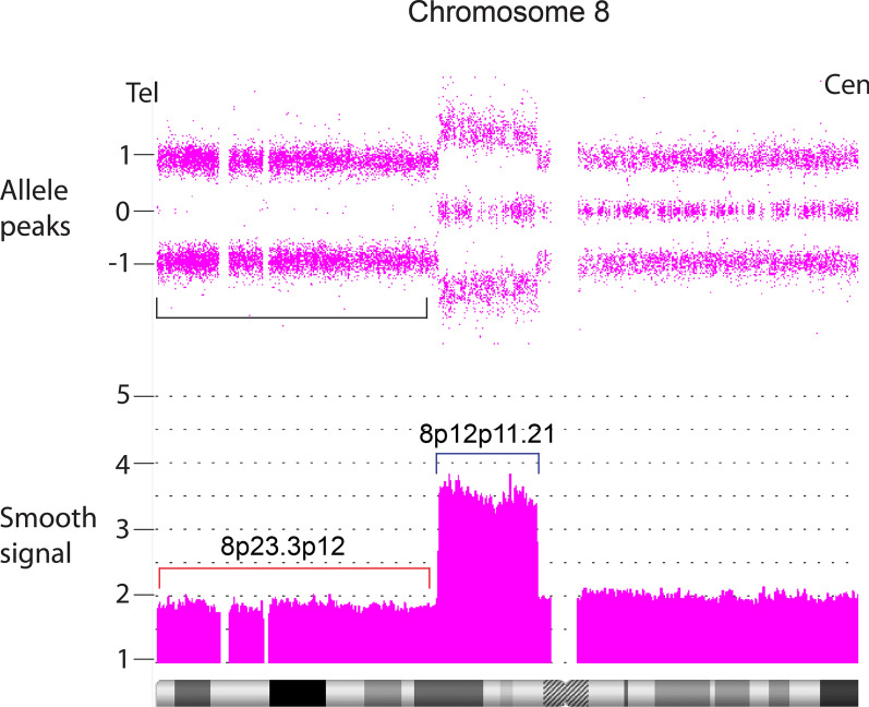
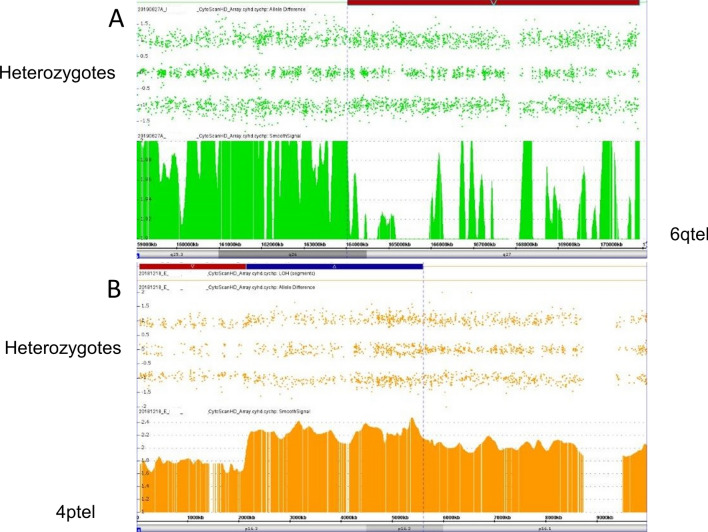
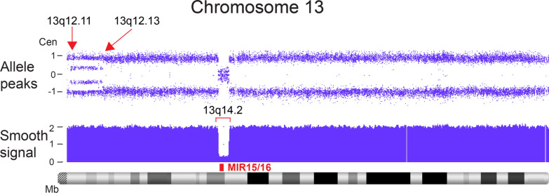
Whole chromosome uniparental disomy (UPD) has been well documented with mechanisms largely understood. However, the etiology of segmental limited UPD (segUPD) is not as clear. In a 10-year period of confirming (> 300) cases of whole chromosome UPD, we identified 86 segmental cases in both prenatal and postnatal samples. Thirty-two of these cases showed mosaic segmental UPD at 11p due to somatic selection associated with Beckwith-Wiedemann syndrome. This study focuses on apparent mechanisms associated with the remaining cases, many of which appear to represent corrections of genomic imbalance such as deletions and derivative chromosomes. In some cases, segmental UPD was associated with the generation of additional genomic imbalance while in others it apparently resulted in restoration of euploidy. Multiple tests utilizing noninvasive prenatal testing (NIPT), chorionic villus sampling (CVS) and amniotic fluid samples from the same pregnancy revealed temporal evidence of correction and a "hotspot" at 1p. Although in many cases the genomic imbalance was dosage "repaired" in the analyzed tissue, clinical effects could be sustained due to early developmental effects of the original imbalance or due to its continued existence in other tissues. In addition, if correction did not occur in the gametes there would be recurrence risks for the offspring of those individuals. Familial microarray allele patterns are presented that differentiate lack of gamete correction from somatic derived gonadal mosaicism. These results suggest that the incidence of segUPD mediated correction is underestimated and may explain the etiology of some clinical phenotypes which are undetected by routine microarray analysis and many exome sequencing studies.

Keywords: Chromosome microarray (CMA); Cytogenetics; Homologous recombination; Mitotic correction; Segmental uniparental disomy; Uniparental disomy.


Conflict of interest statement

Authors PP, CK, SC, SS, and AP are employees of Laboratory Corporation of America with option to hold stock. SH is employed by University of South Florida.

Figures

Fig. 1
Fig. 1
CMA analysis case example of possible segUPD. A > 16 Mb terminal ROH at 5p in case 7 depicted by the purple block with much shorter ROH elsewhere in the analysis inconsistent with a consanguinity correlation. Parental follow-up confirmed maternal segUPD
Fig. 2
Fig. 2
Graphic depiction of mitotic-mediated correction of meiotically derived imbalance. Double-strand break mediated repair results in inter-homologue recombination during S phase of a diploid chromatid (pink) with a chromatid (blue) containing a terminal rearrangement (orange). This results in two chromosome homologues, each containing one recombinant chromatid. Subsequent mitotic segregation can result in two outcomes: two daughter cells that, like the parent cell, are heterozygous for the original imbalance or in the outcome described in the figure. In this case one daughter cell is homozygous for the imbalance and the other is euploid but with uniparental inheritance in the recombinant region, segUPD, detected as a terminal ROH in microarray testing. Survival and expansion are more likely for the euploid daughter cell. However, variable selective pressure, both in cancer and some constitutional alterations, can result in clonal expansion of cells with the imbalance post mitotic recombination
Fig. 3
Fig. 3
Graphic explanation of allele difference dosage plots in the Chromosome Analysis Suite (ChAS)® SNP software. Each SNP at a given genomic position is assigned either an A or B designation (dependent on the polymorphic base pair at that location) with A = 0.5 and B = − 0.5 and the final value equal to the sum of alleles at a particular genomic position. A A heterozygous diploid allele mix consists of either two A alleles (AA) with a value of 1, one A allele and one B allele (AB) with a value of 0, or two B alleles (BB) with a value of -1 that result in the three tracts shown. B A deletion shows either an A or B allele and only two tracts at a value of 0.5 or − 0.5. C A run of copy neutral homozygosity consists of only the AA or BB allele pattern with a value of 1.0 or − 1.0. D A 50:50 mix of two cell lines in which cell line 1 is heterozygous at a certain position and cell line 2 is homozygous at that position. Note that the heterozygous alleles are shifted away from the midline due to the homozygous admixture. E A duplication shows an AAA, AAB, ABB or BBB (4 tract) pattern with an allele difference value of 1.5, 0.5, − 0.5 or − 1.5. F A triplication with 2 identical maternal and paternal copies results in an AAAA, AABB, and BBBB allele pattern with an allele difference value of 2.0, 0 or − 2.0 and three allele tracts. Typical triplications have four or five allele tracts
Fig. 4
Fig. 4
Relative incidence of segUPD subgroups seen during study timeframe
Fig. 5
Fig. 5
Correction of der(1)t(1;17) in case 1. A Partial karyotype of the 16 week amniocyte analysis in case 1. FISH and parental studies confirmed a de novo unbalanced derivative (1)t(1;17)(p36.3;q21) (arrow). B, C Blood array analysis at age 9 showed a terminal 9.4 Mb copy neutral ROH on chromosome 1 initiating at band p36.22 (arrow) with no deletion of 1p or evidence of partial trisomy 17
Fig. 6
Fig. 6
Possible inversion recombinant correction in case 3. A NIPT study, referred due to a cardiac lesion, showing a terminal deletion of 1p and a duplication of terminal 1q (A), with both equivalent to the 13% fetal fraction, consistent with non-mosaic fetal alterations. B Subsequent microarray analysis at 23 weeks of gestation showed no dosage changes of chromosome 1, although a 21.58 Mb terminal 1p ROH was present (B)
Fig. 7
Fig. 7
Novel correction of a derivative 4 in case 6. A Karyotype of the mosaic derivative(4)t(3;4)(p22;q35) still present in 5% of the patient lymphocytes as an adult. B. Whole genome view of ROH greater than 1 Mb from buccal cell CMA. C Isolated view of 3p terminal ROH. SegUPD confirmation is in Additional file 1: Figure S2
Fig. 8
Fig. 8
Apparent mechanism for segUPD 3p in case 6. Mitotic recombination at 3p22 and distal fission at 4q results in loss of the 3p segment from the derivative 4 and segUPD for 3p
Fig. 9
Fig. 9
Incomplete correction of the translocation derivative 21 in case 35. A Partial karyotype showing mosaic cell lines in case 35. Normal cell line (top panel) and derivative (21)t(12;21)(p11.22;q22.2) cell line (bottom panel, arrow). B CMA of chromosome 12 shows a mosaic terminal duplication (bracket) of 30.51 Mb in ~ 60% of cells. C CMA of chromosome 21 shows a mosaic terminal deletion (smooth signal track, bracket) of 7.12 in ~ 60% of cells. Arrow shows the initiation site of mitotic recombination at 21q21.1, resulting in replacement of the der(21) with a segment from the normal homologue and the obligate homozygotic allele dosage. D A single cell in which the der(21) initiates mitotic recombination with the normal homologue. Segregation and selection results in a second cell line with a normal copy number for chromosome 12 and 21, resulting in segUPD 21q21.1->qter
Fig. 10
Fig. 10
Analysis of prenatal deletion in case 9. A Case 9 showing a deletion of 10q present in all cells from a CVS chromosome analysis (top panel, arrow) that was confirmed by a region-specific FISH probe (bottom panel, arrow). B Post-delivery blood CMA revealing a copy neutral terminal ROH on chromosome 10 initiating at band q26.13 (bracket). Confirmation of segUPD is shown in Additional file 1: Figure S3
Fig. 11
Fig. 11
CVS and AF CMAs in case 2. 1p36.22 deletion in CVS analysis (top panel) with a correction to apparent segUPD in the AF analysis (bottom panel). The close proximity of the AF ROH correction initiation site to the original deletion site is consistent with a recombination event in close proximity to the deletion breakpoint
Fig. 12
Fig. 12
Mosaic segUPD15 associated with 3 distinct corrections of an interstitial deletion detected in case 30. Three deletion repair cell lines initiate at different mitotic recombination sites (arrows). The deletion (bracket) is still present in ~ 25% of cells. Note that the allele difference tract shows ~ 75% of cells with homozygosity in the location of the deletion, consistent with the percentage of deletion correction
Fig. 13
Fig. 13
Evidence for somatic origin of mosaic segUPD 12 in case 24. Mosaic apparent segUPD12 detected in the blood of a newborn (top) and, to a lesser extent, in a buccal swab (middle), but absent in the placenta (bottom) in case 24
Fig. 14
Fig. 14
Temporal prenatal analyses in case 4. A NIPT analysis showing an 8.72 Mb terminal deletion (red bracket) contiguous with a 3.44 Mb duplication that extends to 1p36.22 (blue bracket). B Amniocyte CMA revealing a terminal 16.32 Mb copy neutral ROH initiating at band 1p36.13 (lower panel). Pooled placental analysis (top panel) showing ~ 80% mosaicism for the alterations initiating at 1p36.22. Note that the ROH initiates proximally to the duplication
Fig. 15
Fig. 15
CMA and FISH analyses of a case of segUPD associated with a triplication. A Microarray image of case 38, shows 3.56 Mb triplication (blue bracket) of bands p36.13p36.12 on chromosome 1 with an exclusive 2:2 (AABB) heterozygote allele pattern and a contiguous copy neutral terminal 19.35 Mb ROH (bracket). B A dual target interphase FISH image showing triplication of 1p (bracket) with inverted orientation of the middle segment (arrow)
Fig. 16
Fig. 16
Postulated mechanism of segUPD associated with triplications. After passage of a meiotic derived inverted duplication with a contiguous terminal deletion to the zygote, there is somatic rescue by the normal homologue, initiating from the most distal sequence of the inverted duplication. This results in adding a third copy to the duplication and exclusive AABB heterozygote alleles in the CMA (rather than three or four tracts), while correcting the deletion imbalance with terminal segUPD
Fig. 17
Fig. 17
Mosaic example of a segUPD/triplication correction. The fetal demise from patient 46 showed terminal homozygotic alleles and smooth signal dosage consistent with ~ 80% replacement of a terminal 8p deletion/proximal duplication with a copy of the terminal segment from the normal homologue, resulting in terminal segUPD visualized by a terminal ROH (bracket) and a contiguous triplication with the characteristic exclusive balanced AABB heterozygote alleles (smooth signal track, blue bracket). Replacement of the deletion with material from the normal homologue appears to have initiated at the distal end of the contiguous duplication
Fig. 18
Fig. 18
Study showing post zygotic origin of gonadal mosaicism. A Maternal buccal CMA follow-up from the mother of two 6q- offspring revealing a mosaic ~ 11% deletion, red bar. B Maternal buccal follow-up from the mother of two offspring with a 4p deletion-duplication showing ~ 25% deletion (red bar) and duplication (blue bar). Note that the presence of heterozygote alleles in the allele difference tract in the deletion interval are consistent with a post zygotic origin of the gonadal mosaicism and inconsistent with segUPD related correction
Fig. 19
Fig. 19
Example of multiple sites of mitotic recombination in clonal based selection in cancer. Archival sample of a leukemia case showing acquired regions of expanding homozygosity referred to as copy neutral loss of heterozygosity (CN-LOH). Two mosaic contiguous regions of CN-LOH (arrows) on chromosome 13, consistent with the evolution of two cells lines with mitotic recombination mediated homozygosity initiated at 13q12.11 and 13q12.13. The selection of recombinant cells is driven by conversion of a heterozygous deletion of the MIR15/16 tumor suppressor genes, red rectangle) at13q14.2 to homozygosity (bracket). A similar pattern is seen in the conversion to normal in case 30 (Fig. 12)

Similar articles

Cited by

References

    1. Engel E. A new genetic concept: uniparental disomy and its potential effect, isodisomy. Am J Med Genet. 1980;6(2):137–143. doi: 10.1002/ajmg.1320060207. - DOI - PubMed
    1. Papenhausen P, Schwartz S, Risheg H, Keitges E, Gadi I, Burnside RD, Jaswaney V, Pappas J, Pasion R, Friedman K, Tepperberg J. UPD detection using homozygosity profiling with a SNP genotyping microarray. Am J Med Genet A. 2011;155(4):757–768. doi: 10.1002/ajmg.a.33939. - DOI - PubMed
    1. Kearney HM, Kearney JB, Conlin LK. Diagnostic implications of excessive homozygosity detected by SNP-based microarrays: consanguinity, uniparental disomy, and recessive single-gene mutations. Clin Lab Med. 2011;31(4):595–613. doi: 10.1016/j.cll.2011.08.003. - DOI - PubMed
    1. Lapunzina P, Monk D. The consequences of uniparental disomy and copy number neutral loss-of-heterozygosity during human development and cancer. Biol Cell. 2011;103(7):303–317. doi: 10.1042/BC20110013. - DOI - PubMed
    1. Schollen E, Grünewald S, Keldermans L, Albrecht B, Körner C, Matthijs G. CDG-Id caused by homozygosity for an ALG3 mutation due to segmental maternal isodisomy UPD3 (q21. 3-qter) Eur J Med Genet. 2005;48(2):153–158. doi: 10.1016/j.ejmg.2005.01.002. - DOI - PubMed

LinkOut - more resources