. 2021 Jul 20;28(1):16.
doi: 10.1186/s40709-021-00148-3.

PTD-mediated delivery of α-globin chain into Κ-562 erythroleukemia cells and α-thalassemic (HBH) patients' RBCs ex vivo in the frame of Protein Replacement Therapy

Affiliations

PTD-mediated delivery of α-globin chain into Κ-562 erythroleukemia cells and α-thalassemic (HBH) patients' RBCs ex vivo in the frame of Protein Replacement Therapy

Androulla N Miliotou et al. J Biol Res (Thessalon). .

Abstract

Background: α-Thalassemia, a congenital hemoglobinopathy, is characterized by deficiency and/or reduced levels of α-globin chains in serious forms of α-thalassemia (HbH disease/Hb Bart's). This research work deals with a Protein Replacement Therapy approach in order to manage α-thalassemia manifestations, caused by the excess of β-globin chain into HbH RBCs. The main goal was to produce the recombinant human α-globin chain in fusion with TAT, a Protein Transduction Domain, to ex vivo deliver it into HbH patients RBCs, to replace the endogenous missing α-globin chain.

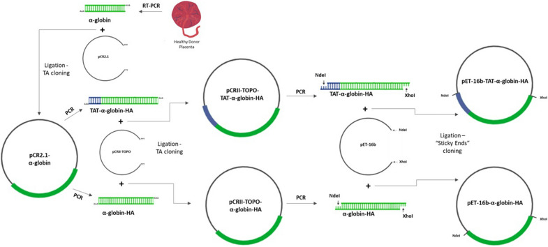
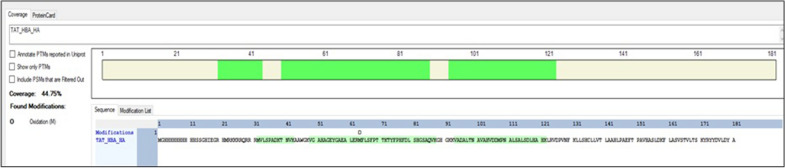
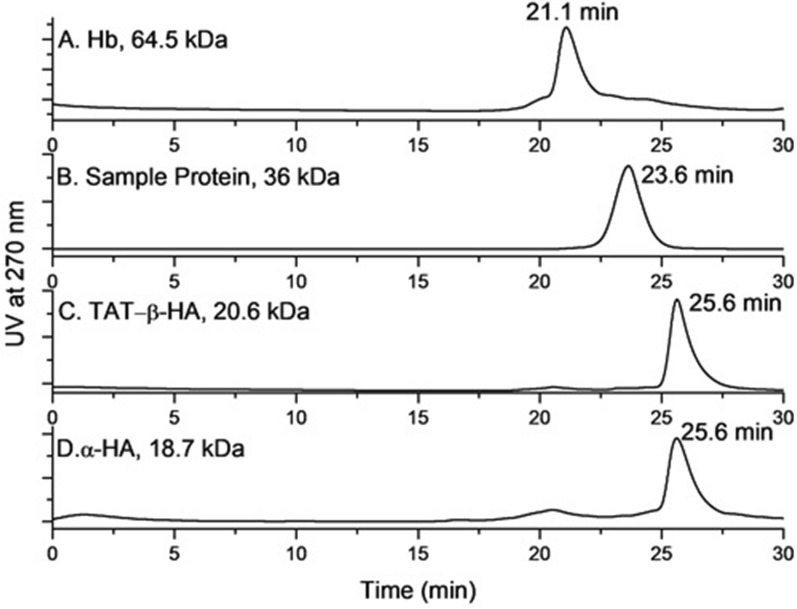
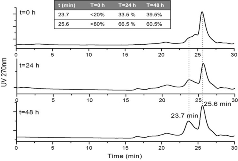
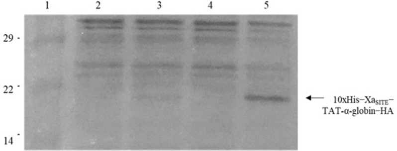
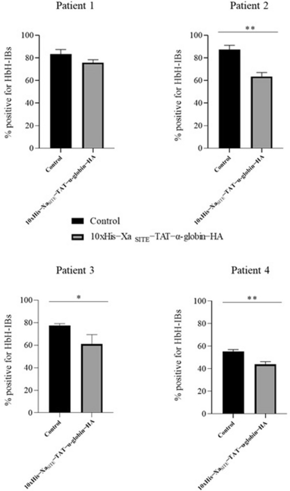
Results: Cloning of the α-globin coding sequence, fused to the nucleotide sequence of TAT peptide was conducted and the human recombinant fusion proteins, 10xHis-XaSITE-α-globin-HA and 10xHis-XaSITE-TAT-α-globin-HA were produced. The ability of human recombinant 10xHis-XaSITE-α-globin-HA to interact in vitro with the previously produced 10xHis-XaSITE-TAT-β-globin-HA and form α-/β-globin heterodimers, was assessed and confirmed by size exclusion chromatography. The recombinant 10xHis-XaSITE-TAT-α-globin-HA was successfully delivered into human proerythroid K-562 cells, during the preliminary transduction evaluation experiments. Finally, the recombinant, TAT-fused α-globin was successfully transduced into RBCs, derived from HbH patients and reduced the formation of HbH-Inclusion Bodies, known to contain harmful β4-globin chain tetramers.

Conclusions: Our data confirm the successful ex vivo transduction of recombinant α-globin chains in HbH RBCs to replace the missing a-globin chain and reduce the HbH-inclusion bodies, seen in α-thalassemias. These findings broaden the possibility of applying a Protein Replacement Therapy approach to module sever forms of α-thalassemia, using recombinant α-globin chains, through PTD technology.

Keywords: HbH thalassemic patients’ RBCs; Intracellular transduction via PTD; LC − MS/MS analysis; Size exclusion chromatography; TAT-α-globin.


Conflict of interest statement

All authors declare no conflict of interests.

Figures

Fig. 1
Fig. 1
Schematic illustration of the cloning procedure, starting with RT-PCR from placenta tissue, derived from a healthy individual, followed by TA cloning of the a-globin CDS and then TA cloning of amplified TAT-α-globin-HA and α-globin-HA into pCRII-TOPO vector. Then, «sticky ends» cloning was conducted, with the restriction enzymes NdeI and XhoI, to generate the recombinant expression vectors pET-16b-TAT-α-globin-HA and pET-16b-α-globin-HA
Fig. 2
Fig. 2
Western Blot of recombinant protein extracts, separated on 15% SDS-PAGE, transferred and immunoblotted with anti-His.IgG. A The bacterial-IBs’ protein extracts (in 1 M l-Arg, pH 8.0). Lane 1: protein molecular mass marker; Lane 2: 10xHis-XaSITE-TAT-α-globin-HA (~ 20.2 kDa); Lane 3: protein molecular mass marker; Lane 4: 10xHis-XaSITE-α-globin-HA (~ 18.7 kDa); B The soluble-purified by Ni2+-NTA chromatography-recombinant proteins, eluted with 10 mM imidazole. Lane 1: protein molecular mass marker; Lane 2: 10xHis-XaSITE-TAT-α-globin-HA (~ 20.2 kDa); Lane 3: 10xHis-XaSITE-α-globin-HA (~ 18.7 kDa). Higher bands at 29—41 kDa indicate the corresponding protein dimers, while lower bands may indicate proteolysis products
Fig. 3
Fig. 3
Retrieval of recombinant α-globin fusion protein variants purified by affinity Ni2+-NTA chromatography under denaturing conditions and analyzed by 15% SDS − PAGE and Coomassie Blue staining: A SDS-PAGE analysis of the purification of 10xHis-XaSITE-α-globin-HA protein (~ 18.7 kDa). Lane 1: Crude extract of induced bacterial cells transformed by pET-16b-α globin-HA; Lane 2: Flow-through fraction of the Ni2+-NTA affinity column; Lane 3: Wash Buffer extract with 10 mM imidazole; Lane 4: Elution with 20 mM imidazole; Lane 5: Elution with 40 mM imidazole; Lane 6: protein molecular mass marker; Lane 7: Elution with 100 mM imidazole; Lane 8: Elution with 200 mM imidazole; Lane 9: Elution with 400 mM imidazole. B SDS-PAGE analysis of the purification of 10xHis-XaSITE-TAT-α-globin-HA protein (~ 20.2 kDa). Lane 1: Crude extract of induced bacterial cells transformed by pET-16b-TAT-α globin-HA; Lane 2: Flow-through fraction of the Ni2+-NTA affinity column; Lane 3: Wash Buffer extract with 10 mM imidazole; Lane 4: Elution with 20 mM imidazole; Lane 5: Elution with 40 mM imidazole; Lane 6: Elution with 100 mM imidazole; Lane 7: Elution with 200 mM imidazole; Lane 8: Elution with 400 mM imidazole; Lane 9: protein molecular mass marker
Fig. 4
Fig. 4
Identification of recombinant 10xHis-XaSITE-TAT-α-globin-HA (TAT_HBA_HA) protein construct by LC − MS/MS. The highlighted peptides confidently identified cover 44.75% of the theoretical construct sequence
Fig. 5
Fig. 5
Size exclusion HPLC chromatograms: A hemoglobin tetramer (64.5 kDa) (tR = 21.1 min); B soluble (in PBS 1 ×) sample protein (∼36 kDa) (tR = 23.628 min); C soluble monomer 10xHis-XaSITE-TAT-β-globin-HA (∼20.6 kDa), prior incubated with DTT for 48 h (tR = 25.6 min); D soluble monomer 10xHis-XaSITE-α-globin-HA (∼18.7 kDa), prior incubated with DTT for 48 h (tR = 25.6 min)
Fig. 6
Fig. 6
Size exclusion HPLC chromatograms of reaction mixtures of α- and β-monomers at t = 0 h, t = 24 h and t = 48 h
Fig. 7
Fig. 7
Western Blot of electrophoresed cell lysates derived from K-562 cells, transduced with 50 μg ml−1 soluble 10xHis − XaSITE − TAT − α-globin − HA, immunoblotted using anti-HA.IgG. Lane 1: protein molecular mass marker; Lane 2: lysate from control (untreated) cells, 30 min; Lane 3: lysate from cells transduced with 10xHis − XaSITE − TAT − α-globin − HA, incubated for 30 min.; Lane 4: lysate from control (untreated) cells, 1 h; Lane 5: lysate from cells transduced with 10xHis − XaSITE − TAT − α-globin − HA, incubated for 1 h
Fig. 8
Fig. 8
Western Blot of electrophoresed cell lysates derived from HbH patients’ peripheral RBCs (Patient 3), transduced with bacterial-IBs, enriched in 10xHis-XaSITE-TAT-α-globin-HA, immunoblotted using anti-His.IgG and anti-GAPDH.IgG. Patient 3: Lane 1: protein molecular mass marker; Lane 2: lysate from control (untreated) cells, 48 h; Lane 3: lysate from cells incubated with 1 M l-Arg, for 48 h; Lanes 4–6: lysate from cells transduced with bacterial-IBs, enriched in 10xHis-XaSITE-TAT-α-globin-HA, incubated for 48 h
Fig. 9
Fig. 9
Methylene violet staining for the evaluation of HbH-IBs’ reduction, in HbH RBCs upon incubation with 10xHisXaSITE-TAT-α-globin-HA. Patient 2: A Untreated HbH patient’s RBCs, and B HbH patient’s RBCs incubated with IBs, enriched in 10xHis-XaSITE-TAT-α-globin-HA, for 48 h. Patient 3: A Untreated HbH patient’s RBCs, B HbH patient’s RBCs incubated with 1 M l-Arg, for 48 h, C HbH patient’s RBCs incubated with IBs, enriched in 10xHis-XaSITE-TAT-α-globin-HA, for 48 h
Fig. 10
Fig. 10
Diagrams for % positive for HbH-IBs in RBCs, derived from HbH patients (Patients 1–4), which were reduced after incubation with 10xHis-XaSITE-TAT-α-globin-HA, for 48 h

Similar articles

Cited by

References

    1. Papadopoulou LC, Tsiftsoglou AS. Transduction of human recombinant proteins into mitochondria as a protein therapeutic approach for mitochondrial disorders. Pharm Res. 2011;28(11):2639–2656. doi: 10.1007/s11095-011-0546-y. - DOI - PubMed
    1. Brasseur R, Divita G. Happy birthday cell penetrating peptides: already 20 years. Biochim Biophys Acta. 2010;1798(12):2177–2181. doi: 10.1016/j.bbamem.2010.09.001. - DOI - PubMed
    1. Vives E. Present and future of cell-penetrating peptide mediated delivery systems: "is the Trojan horse too wild to go only to Troy?". J Control Release. 2005;109(1–3):77–85. doi: 10.1016/j.jconrel.2005.09.032. - DOI - PubMed
    1. Snyder EL, Dowdy SF. Cell penetrating peptides in drug delivery. Pharm Res. 2004;21(3):389–393. doi: 10.1023/B:PHAM.0000019289.61978.f5. - DOI - PubMed
    1. Eguchi A, Dowdy SF. siRNA delivery using peptide transduction domains. Trends Pharmacol Sci. 2009;30(7):341–345. doi: 10.1016/j.tips.2009.04.009. - DOI - PubMed

Grants and funding

LinkOut - more resources