Review
. 2022 Jan 1;28(1):5-12.
doi: 10.1158/1078-0432.CCR-21-1193. Epub 2021 Jul 20.

The Liver-Immunity Nexus and Cancer Immunotherapy

Affiliations
Review

The Liver-Immunity Nexus and Cancer Immunotherapy

James C Lee et al. Clin Cancer Res. .

Abstract

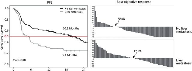
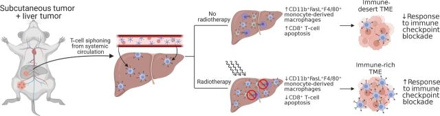
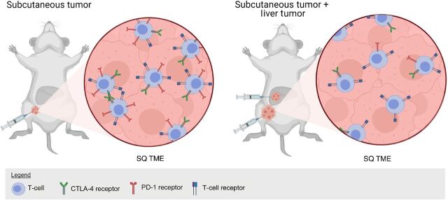
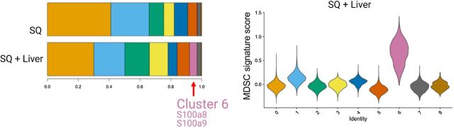
The impact of liver metastases on immune checkpoint-inhibitor effectiveness in patients with solid-tumor malignancies has been the focus of several recent clinical and translational studies. We review the literature describing the immune functions of the liver and particularly the mechanistic observations in these studies. The initial clinical observation was that pembrolizumab appeared to be much less effective in melanoma and non-small cell lung cancer (NSCLC) patients with liver metastasis. Subsequently other clinical studies have extended and reported similar findings with programmed death-1 (PD-1) and programmed death ligand-1 (PD-L1) inhibitors in many cancers. Two recent translational studies in animal models have dissected the mechanism of this systemic immune suppression. In both studies CD11b+ suppressive macrophages generated by liver metastasis in a two-site MC38 model appear to delete CD8+ T cells in a FasL-dependent manner. In addition, regulatory T-cell (Treg) activation was observed and contributed to the distal immunosuppression. Finally, we discuss some of the interventions reported to address liver immune suppression, such as radiation therapy, combination checkpoint blockade, and Treg depletion.


Figures

Figure 1. Kaplan–Meier OS curves for melanoma patients without (black line) and with liver metastasis (gray line) and waterfall response curves for melanoma without and with liver metastasis. All patients were treated with pembrolizumab immunotherapy as described in Tumeh and colleagues (32). Figure originally published in Cancer Immunology Research (32).
Figure 1.
Kaplan–Meier OS curves for melanoma patients without (black line) and with liver metastasis (gray line) and waterfall response curves for melanoma without and with liver metastasis. All patients were treated with pembrolizumab immunotherapy as described in Tumeh and colleagues (32). Figure originally published in Cancer Immunology Research (32).
Figure 2. Schematic diagram showing experimental protocol for mice with subcutaneous + liver-tumor treated with no radiotherapy or with radiotherapy showing changes in the subcutaneous tumor as depicted in Yu and colleagues (37). Figure created with BioRender.com.
Figure 2.
Schematic diagram showing experimental protocol for mice with subcutaneous + liver-tumor treated with no radiotherapy or with radiotherapy showing changes in the subcutaneous tumor as depicted in Yu and colleagues (37). Figure created with BioRender.com.
Figure 3. Effect of radiotherapy on immune-cell populations in the liver. Large plot; viSNE analysis of CyTOF immunophenotyping of livers from mice with both subcutaneous and liver tumors, treated with IgG, anti–PD-L1, liver-directed radiotherapy, or combination therapy, as depicted in Yu and colleagues (37). Figure previously published in Nature Medicine (37); reprinted with permission.
Figure 3.
Effect of radiotherapy on immune-cell populations in the liver. Large plot; viSNE analysis of CyTOF immunophenotyping of livers from mice with both subcutaneous and liver tumors, treated with IgG, anti–PD-L1, liver-directed radiotherapy, or combination therapy, as depicted in Yu and colleagues (37). Figure previously published in Nature Medicine (37); reprinted with permission.
Figure 4. Liver metastasis induces systemic loss of antigen-specific T cells. Immunofluorescent staining of CD8+ cells in MC38 subcutaneous tumors from mice with subcutaneous tumors only or with subcutaneous and liver tumors. Analysis was done at 10 days after therapy initiation. Figure previously published in Nature Medicine (37); reprinted with permission.
Figure 4.
Liver metastasis induces systemic loss of antigen-specific T cells. Immunofluorescent staining of CD8+ cells in MC38 subcutaneous tumors from mice with subcutaneous tumors only or with subcutaneous and liver tumors. Analysis was done at 10 days after therapy initiation. Figure previously published in Nature Medicine (37); reprinted with permission.
Figure 5. Mouse model with MC38 colon cancer syngeneic tumor implanted into the subcutaneous tissue or subcutaneous and liver as depicted in Lee JC and colleagues (54). In liver and subcutaneous tumor-bearing mice, there are reduced PD-1hi/CTLA-4hi CD8+ T cells, and these tumors have reduced response to PD-1 blockade. Figure created with BioRender.com.
Figure 5.
Mouse model with MC38 colon cancer syngeneic tumor implanted into the subcutaneous tissue or subcutaneous and liver as depicted in Lee JC and colleagues (54). In liver and subcutaneous tumor-bearing mice, there are reduced PD-1hi/CTLA-4hi CD8+ T cells, and these tumors have reduced response to PD-1 blockade. Figure created with BioRender.com.
Figure 6. Single-cell RNA sequencing in Lee and colleagues (54) showed a myeloid population consistent with an MDSC phenotype in liver MC38 tumor mice, cluster 6 shown in the histogram and in violin plots showing relative MDSC score ordered by monocyte/myeloid cell subclusters. Figure previously published in Science Immunology (54); reprinted with permission.
Figure 6.
Single-cell RNA sequencing in Lee and colleagues (54) showed a myeloid population consistent with an MDSC phenotype in liver MC38 tumor mice, cluster 6 shown in the histogram and in violin plots showing relative MDSC score ordered by monocyte/myeloid cell subclusters. Figure previously published in Science Immunology (54); reprinted with permission.

References

    1. Juza RM, Pauli EM. Clinical and surgical anatomy of the liver: a review for clinicians. Clin Anat 2014;27:764–9. - PubMed
    1. Andersson ER. In the zone for liver proliferation. Science 2021;371:887–8. - PubMed
    1. He L, Pu W, Liu X, Zhang Z, Han M, Li Y, et al. Proliferation tracing reveals regional hepatocyte generation in liver homeostasis and repair. Science 2021;371:eabc4346. - PubMed
    1. Wei Y, Wang YG, Jia Y, Li L, Yoon J, Zhang S, et al. Liver homeostasis is maintained by midlobular zone 2 hepatocytes. Science 2021;371:eabb1625. - PMC - PubMed
    1. Calne RY, Sells RA, Pena JR, Davis DR, Millard PR, Herbertson BM, et al. Induction of immunological tolerance by porcine liver allografts. Nature 1969;223:472–6. - PubMed

Publication types