. 2021 Jul 20;12(1):4399.
doi: 10.1038/s41467-021-24490-1.

Miniature neurotransmission is required to maintain Drosophila synaptic structures during ageing

Affiliations

Miniature neurotransmission is required to maintain Drosophila synaptic structures during ageing

Soumya Banerjee et al. Nat Commun. .

Abstract

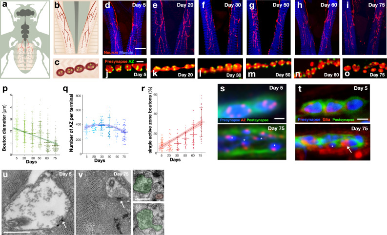
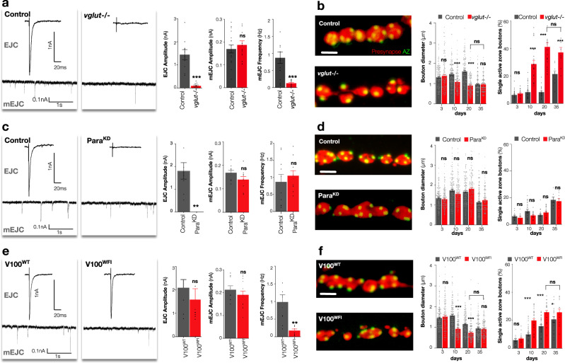
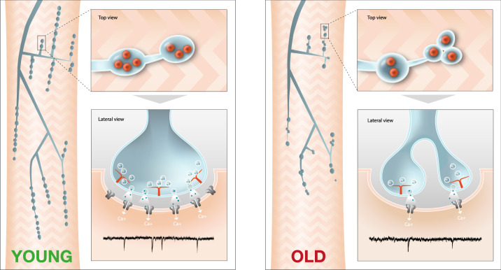
The decline of neuronal synapses is an established feature of ageing accompanied by the diminishment of neuronal function, and in the motor system at least, a reduction of behavioural capacity. Here, we have investigated Drosophila motor neuron synaptic terminals during ageing. We observed cumulative fragmentation of presynaptic structures accompanied by diminishment of both evoked and miniature neurotransmission occurring in tandem with reduced motor ability. Through discrete manipulation of each neurotransmission modality, we find that miniature but not evoked neurotransmission is required to maintain presynaptic architecture and that increasing miniature events can both preserve synaptic structures and prolong motor ability during ageing. Our results establish that miniature neurotransmission, formerly viewed as an epiphenomenon, is necessary for the long-term stability of synaptic connections.


Conflict of interest statement

The authors declare no competing interests.

Figures

Fig. 1
Fig. 1. Drosophila synaptic terminal structures fragment, neurotransmission declines and motor behaviour is diminished during ageing.
Schematic of adult Drosophila HB9 labelled motor neurons (a, red) which innervate a subset of ventral and dorsal muscles of the anterior abdomen including A2 mvim (b) producing terminal bouton varicosities (c) with multiple active zones of synaptic vesicle release (yellow). di Representative images of progressive degeneration of HB9 abdominal synaptic terminals (red, GFP) innervating A2 abdominal muscles (blue, phalloidin) from 5 days after eclosion to 75 days. Magnification is identical for all images. jo Representative images of progressive fragmentation of synaptic boutons (red, GFP) through reduction in size and number of active zones (green, Brp) per bouton during ageing. Magnification is identical for all images. p Quantification of diminishment of bouton diameters during ageing. q Quantification of the number of active zones during ageing. r Quantification of bouton fragmentation during ageing as measured by the percentage of boutons with only a single active zone. s Representative images of presynaptic boutons (blue, mCherry) at day 5 (upper panel) and day 75 (lower panel). Boutons are labeled with the active zone marker Brp (red) and the postsynaptic membrane marker Dlg (green). Asterisks indicate fragmentation of presynaptic terminal at day 75. Magnification is identical for both images. t Example of postsynaptic membrane (Green, Dlg) becoming opposed by peripheral glia (Red, mCherry) [indicated by arrow] as presynaptic terminals fragment (indicated by asterisk) (Blue, GFP) in 75-day old  animals (lower panel), which is not found in 5-day-old young animals (upper panel). Magnification is identical for both images. n = 4 biologically independent preparations. u, v Representative micrographs of bouton at day 5 and day 75, respectively. v (Right panels) serial micrograph shows an example of a fragmented bouton which is associated with other boutons by a thin process. Arrows indicate T-bars. n = 11 (u) and 5 (v) biologically independent preparations. One-way ANOVA, ***p < 0.001, R2 = 0.28, n = 150 boutons per timepoint (p), ***p < 0.001, R2 = 0.11, n ≥ 21presynaptic terminals per timepoint (q), ***p < 0.001, R2 = 0.48, n ≥ 27 presynaptic terminals per timepoint (r), Tukey’s multiple comparison tests were used to compare the mean of each timepoint with the mean of all other timepoints (ns = not significant, *p ≤ 0.033, **p ≤ 0.002, ***p ≤ 0.001). All statistical analysis details with a precise value of ‘n’ are reported in Supplementary Table 2 and in Source Data file. Scale bars, 40 μm (di), 2 μm (jo, s, t), 500 nm (u, v), 250 nm (v, right panel). Data are represented as mean ± SD. n = biologically independent samples.
Fig. 2
Fig. 2. Neurotransmission declines and motor behaviour is diminished during ageing.
a, b Representative traces of excitatory junctional currents (EJCs) (above) and miniature excitatory junctional currents (mEJCs) (below) from 5-day-old and 75-day-old A2 mvim  terminals. c Quantification of EJC amplitude decline during ageing. d Quantification of mEJC amplitude stability during ageing. e Quantification of mEJC frequency decline during ageing. f Quantification of declining motor ability during ageing. g Summary diagram indicates the progressive change of key ageing parameters as a percentage of the maximum observed phenotype. Compared to young animals (day 5) individual timepoints were significantly different at middle age (day 30) for increased bouton fragmentation, early old age (day 60) for EJC amplitude, early old age (day 60) for mEJC frequency and middle age (day 30) for motor ability. One-way ANOVA, **p = 0.004, R2 = 0.31, n ≥ 7 presynaptic terminals per timepoint (c), *p = 0.04, R2 = 0.11, n ≥ 13 presynaptic terminals per timepoint (d), **p = 0.004, R2 = 0.15, n ≥ 13 presynaptic terminals per timepoint (e), ***p < 0.001, R2 = 0.86, n ≥ 5 groups per timepoint (f), Tukey’s multiple comparison tests were used to compare the mean of each timepoint with the mean of all other timepoints (ns = not significant, *p ≤ 0.033, **p ≤ 0.002, ***p ≤ 0.001). All statistical analyses with a precise value of ‘n’ are reported in Supplementary Table 2 and in Source Data file. Data are represented as mean ± SD. n = biologically independent samples.
Fig. 3
Fig. 3. Miniature but not evoked neurotransmission is required for adult synapse maintenance.
a Representative EJC (above) and mEJC (below) traces, quantification of EJC amplitude, mEJC amplitude and frequency of control [UAS > GFP/+; HB9 > Gal4, Tub > Gal80ts] and HB9 neuron selective adult conditional vglut−/− mutants [Df(2R)371/B3RT_vglut_B3RT; HB9 > Gal4, UAS > GFP, Tub > Gal80ts/UAS > B3] at 20 days after eclosion. b Representative images of presynaptic HB9 neuron boutons (red, GFP) and active zones (Brp, green), quantification of bouton diameters and percentage of boutons with only a single active zone (bouton fragmentation) of control and vglut mutants until 35 days after eclosion. c Representative EJC and mEJC traces, quantification of EJC amplitude, mEJC amplitude and frequency of control [UAS > luciferaseRNAi/HB9 > Gal4, UAS > GFP, Tub > Gal80ts] and HB9 neuron selective adult conditional Para knockdown mutants (ParaKD) [UAS > GFP/+; HB9 > Gal4, Tub > Gal80ts/UAS > ParaRNAi] at 20 days after eclosion. d Representative images of presynaptic HB9 neuron boutons and active zones, quantification of bouton diameters and bouton fragmentation as measured by the percentage of boutons with only a single active zone of control and ParaKD mutants until 35 days after eclosion. e Representative EJC and mEJC traces, quantification of EJC amplitude, mEJC amplitude and frequency of HB9 neuron selective adult conditional V100WT [UAS > V1003′UTR_RNAi/UAS > V100WT; HB9 > Gal4, UAS > GFP, TubGal80ts] and V100WFI mutants [UAS > V1003UTR_RNAi/UAS > V100WFI; HB9 > Gal4, UAS > GFP, TubGal80ts] at 20 days after eclosion. f Representative images of presynaptic HB9 neuron boutons and active zones, quantification of bouton diameters and percentage of boutons with only a single active zone of V100WT and V100WFI mutants until 35 days after eclosion. Unpaired two-tailed t-test, ***p < 0.001, n ≥ 9 presynaptic terminals per timepoint (a, EJC amplitude), p = 0.249, n ≥ 17 presynaptic terminals per timepoint (a, mEJC amplitude), ***p < 0.001, n ≥ 17 presynaptic terminals per timepoint (a, mEJC frequency), ***p < 0.001, n ≥ 6 presynaptic terminals per timepoint (c, EJC amplitude), p = 0.09, n ≥ 10 presynaptic terminals per timepoint (c, mEJC amplitude), p = 0.5, n ≥ 10 presynaptic terminals per timepoint (c, mEJC frequency), p = 0.4, n ≥ 8 presynaptic terminals per timepoint (e, EJC amplitude), p = 0.06, n ≥ 8 presynaptic terminals per timepoint (e, mEJC amplitude), **p = 0.004, n ≥ 8 presynaptic terminals per timepoint (e, mEJC frequency). Two-way ANOVA, followed by Sidak’s multiple comparison tests were used to compare the mean of control and experimental genotypes in that timepoint, ns = not significant, ***p ≤ 0.001, n = 150 boutons per timepoint (b, bouton diameters), ns not significant, ***p < 0.001, n ≥ 9 presynaptic terminals per timepoint (b, single active zone boutons), ns not significant, n = 150 boutons per timepoint (d, bouton diameters), ns not significant, n ≥ 9 presynaptic terminals per timepoint (d, single active zone boutons), ns = not significant, ***p ≤ 0.001, n = 150 boutons per timepoint (f, bouton diameters), ns not significant, ***p < 0.001, n ≥ 14 presynaptic terminals per timepoint (f, single active zone boutons). All statistical analysis details with a precise value of ‘n’ are reported in Supplementary Tables 4 and 5 and in Source Data file. Scale bar = 2 μm (b, d, f). Experiments were carried out at 29 °C. Data are represented as mean ± SEM. n = biologically independent samples.
Fig. 4
Fig. 4. Increasing miniature neurotransmission preserves synapses and prolongs motor ability during ageing.
a Representative EJC (above) and mEJC (below) traces, quantification of EJC amplitude, mEJC amplitude and frequency of HB9 neuron selective adult conditional murine Cpx rescued wild type (mCpxWT) [UAS > CpxRNAi/UAS > GFP; HB9 > Gal4, TubGal80ts/cpxSH1, UAS > mCpxWT] and mCpx Helix Breaker’ (mCpxHB) [UAS > CpxRNAi/UAS > GFP; HB9 > Gal4, Tub > Gal80ts/cpxSH1, UAS > mCpxHB] mutants at 20 days after eclosion. b Representative images of presynaptic HB9 neuron boutons (red, GFP) and active zones (Brp, green), quantification of bouton diameters and percentage of boutons with only a single active zone (bouton fragmentation) of mCpxWT and mCpxHB mutants until 40 days after eclosion. Images at same magnification. c Quantification of the climbing ability of motor neuron selective adult conditional mCpx wild-type mCpxWT [UAS > CpxRNAi/OK6 > Gal4, UAS > GFP; Tub>Gal80ts/cpxSH1, UAS > mCpxWT] and mCpxHB [UAS > CpxRNAi/OK6 > Gal4, UAS > GFP; Tub > Gal80ts/cpxSH1, UAS > mCpxHB]. Unpaired two-tailed t-test, p = 0.89, n ≥ 6 presynaptic terminals per timepoint (a, EJC amplitude), p = 0.81, n ≥ 15 presynaptic terminals per timepoint (a, mEJC amplitude), ***p < 0.003, n ≥ 15 presynaptic terminals per timepoint (a, mEJC frequency). Two-way ANOVA, followed by Sidak’s multiple comparison tests were used to compare the mean of control and experimental genotypes in that timepoint, ns not significant, ***p ≤ 0.001, n ≥ 149 boutons per timepoint (b, bouton diameters), ns not significant, **p < 0.002, ***p < 0.001, n ≥ 7 presynaptic terminals per timepoint (b, single active zone boutons), ns not significant, *p < 0.033, ***p < 0.001, n ≥ 4 groups per timepoint (c). All statistical analysis details with a precise value of ‘n’ are reported in Supplementary Tables 4 and 6 and in Source Data file. Scale bar = 2 μm (b). Experiments were carried out at 29 °C. Data are represented as mean ± SEM. n = biologically independent samples.
Fig. 5
Fig. 5. Model of NMJ synaptic structural changes in response to miniature event decline during ageing.
Miniature event frequency declines during ageing. In response, synaptic bouton diameters decrease and there is an increase in the number of small boutons with only a single active zone. All of these observations are consistent with young multiple active zone boutons fragmenting into smaller single active zone boutons as animals age. Reducing miniature events can accelerate this bouton fragmentation process in young animals while increasing miniature event frequency can delay synapse fragmentation in ageing animals.

References

    1. Bano D, Agostini M, Melino G, Nicotera P. Ageing, neuronal connectivity and brain disorders: an unsolved ripple effect. Mol. Neurobiol. 2011;43:124–130. doi: 10.1007/s12035-011-8164-6. - DOI - PubMed
    1. Burke SN, Barnes CA. Neural plasticity in the ageing brain. Nat. Rev. Neurosci. 2006;7:30–40. doi: 10.1038/nrn1809. - DOI - PubMed
    1. Cizeron M, et al. A brainwide atlas of synapses across the mouse life span. Science. 2020;369:270–275. doi: 10.1126/science.aba3163. - DOI - PMC - PubMed
    1. Petralia RS, Mattson MP, Yao PJ. Communication breakdown: the impact of ageing on synapse structure. Ageing Res. Rev. 2014;14:31–42. doi: 10.1016/j.arr.2014.01.003. - DOI - PMC - PubMed
    1. Oda K. Age changes of motor innervation and acetylcholine receptor distribution on human skeletal muscle fibres. J. Neurol. Sci. 1984;66:327–338. doi: 10.1016/0022-510X(84)90021-2. - DOI - PubMed

Publication types

MeSH terms

Substances