. 2021 Jul 20;12(1):4402.
doi: 10.1038/s41467-021-24712-6.

Dysfunction of the key ferroptosis-surveilling systems hypersensitizes mice to tubular necrosis during acute kidney injury

Affiliations

Dysfunction of the key ferroptosis-surveilling systems hypersensitizes mice to tubular necrosis during acute kidney injury

Wulf Tonnus et al. Nat Commun. .

Abstract

Acute kidney injury (AKI) is morphologically characterized by a synchronized plasma membrane rupture of cells in a specific section of a nephron, referred to as acute tubular necrosis (ATN). Whereas the involvement of necroptosis is well characterized, genetic evidence supporting the contribution of ferroptosis is lacking. Here, we demonstrate that the loss of ferroptosis suppressor protein 1 (Fsp1) or the targeted manipulation of the active center of the selenoprotein glutathione peroxidase 4 (Gpx4cys/-) sensitize kidneys to tubular ferroptosis, resulting in a unique morphological pattern of tubular necrosis. Given the unmet medical need to clinically inhibit AKI, we generated a combined small molecule inhibitor (Nec-1f) that simultaneously targets receptor interacting protein kinase 1 (RIPK1) and ferroptosis in cell lines, in freshly isolated primary kidney tubules and in mouse models of cardiac transplantation and of AKI and improved survival in models of ischemia-reperfusion injury. Based on genetic and pharmacological evidence, we conclude that GPX4 dysfunction hypersensitizes mice to ATN during AKI. Additionally, we introduce Nec-1f, a solid inhibitor of RIPK1 and weak inhibitor of ferroptosis.


Conflict of interest statement

A. L., C. S, and M. G issued a patent for Nec-1f with the number 20160943.5. M. C. is co-founder and shareholder of ROSCUE Therapeutics GmbH. Apart from this, all authors declare no conflict of interest regarding any of the presented data in this paper.

Figures
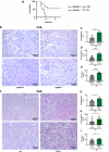
Fig. 1
Fig. 1. Genetic evidence for the contribution of ferroptosis to acute kidney injury.
A Loss of the selenocysteine residue of GPX4 sensitizes mice to acute tubular ferroptosis. Fifthteen days after induction of the Cre-driver, severe renal ischemia-reperfusion injury (IRI) was performed in Gpx4cys/− mice, and overall survival is presented in a Kaplan−Meier plot. B Mice were induced as in (A), and standard IRI was performed. Forty-eight hours after the onset of reperfusion, B representative microphotographs of periodic acid-Schiff (PAS)-stained histological sections of IRI-treated mice are presented and the C tubular damage score was quantified. Serum levels of creatinine (D) and urea (E) were detected. F IRI-treated wild-type C57Bl/6 mice and Fsp1-deficient mice underwent standard renal IRI. Forty-eight hours following the onset of reperfusion, representative PAS-stained histological sections are presented and the tubular damage score was quantified (G). Serum concentrations of creatinine (H) and urea (I) were measured. Bar graphs represent the mean + /− SD. * p < 0.05, *** p < 0.001, ****p < 0.0001.
Fig. 2
Fig. 2. Nec-1f inhibits RIPK1-dependent necroptosis.
A Structures of small molecules used in this study. B Representative co-autoxidations of cumene and STY-BODIPY initiated by Azobisisobutyronitrile (AIBN) and carried out in the presence of Nec-1f or 2,2,7,8-pentamethyl-6-chromanol (PMC). C Representative co-autoxidations of STY-BODIPY and the polyunsaturated fatty acids of egg phosphatidylcholine liposomes initiated by MeOAMVN and carried out in the presence of Nec-1f, PMC, and Fer-1. D Inhibition rate constants (kinh) and stoichiometries (n) derived from the initial rates and lengths of the inhibited periods, respectively, of the data in panel (C). E HT29 cells were treated for 16 h with TNFα (20 ng/ml), birinapant (1 µM), and zVAD-fmk (20 µM) (TSZ) for the induction of necroptosis. The influence of small molecule inhibitors on necroptosis induction is shown by 7-aminoactinomycin (7-AAD) and annexin V positivity. (n = 8 for DMSO, TSZ and Nec-1f; n = 4 for Nec-1s and Fer-1) F HT29 cells were treated for indicated times with TSZ and simultaneous Nec-1f (10 µM) treatment. pMLKL (S358) positivity is shown by western blotting, MLKL and β-actin serve as loading controls. G, H HT29 cells were treated for indicated times with TSZ in the presence of Nec-1s (10 µM), Fer-1 (1 µM) and Nec-1f (10 µM). pMLKL (S358) and pRIPK1 (S166) are demonstrated. I NIH3T3 cells were treated with TZ for 16 h and stained for 7-AAD and annexin V (n = 4). The bar graph shows mean +/− SD. Statistical analysis was performed using one-way ANOVA (post hoc Tukey’s). ** p < 0.01, *** p < 0.001.
Fig. 3
Fig. 3. Nec-1f inhibits ferroptosis.
A HT1080 cells were treated for 6 or 24 h with ferroptosis inducers (FINs) or ferroptocide in the presence of Fer-1 (1 µM), Nec-1s (30 µM) or Nec-1f (30 µM), respectively, as indicated. B NIH3T3 cells were treated as in (A). C NIH3T3 cells were treated for 16 h with either 1.13 µM RSL3 alone, TZ alone, or the combination of RSL3 plus TZ. While Fer-1 or Nec-1s fail to rescue NIH3T3 cells from necrosis, Nec-1f does. Plots show stainings of 7-aminoactinomycin (7-AAD) and annexin V (n = 3). The bar graph shows mean + /− SD. Statistical analysis was performed using one-way ANOVA (post hoc Tukey’s). * p < 0.05, ** p < 0.01.
Fig. 4
Fig. 4. Ferroptosis in primary tubular cells and in freshly isolated murine renal tubules is prevented by ferroptosis inhibitors.
A Still images of a time-lapse video (Supplementary movie 1) of primary murine renal tubular cells demonstrating cell death propagation (SYTOXTM Green) following ferroptosis induction with 1.13 µM RSL3. B The human renal tubular cell line CD10-135 was treated for indicated times with type 1-4 ferroptosis inducers (erastin, RSL3, FIN56, FINO2) or ferroptocide in the presence of Fer-1, Nec-1s, or Nec-1f. Lactate dehydrogenase (LDH) release is presented (n = 3). C Primary kidney tubular cells were treated with RSL3 for 14 h. Note the complete reversal of LDH release by Fer-1 (n = 2). D Primary kidney tubular cells were treated with RSL3 for 14 h in the presence of Nec-1f (n = 3). E Still images and magnifications of Supplementary movie 2 demonstrating characteristic morphological changes (“wave of death”-like cell death progression) in freshly isolated renal tubules that were left unstimulated. F LDH release as a function of time of freshly isolated primary kidney tubules that were left untreated in standard medium (n = 3). G LDH release from unstimulated freshly isolated primary kidney tubules in the presence of 5 mM glycine (DMSO, 0 h, DMSO 2 h and RSL3 2 h n = 6, Fer-1 and Nec-1 n = 3). H Freshly isolated renal tubules were cultured for 2 h in a glycine-containing medium in the presence of RSL3 in the presence of Fer-1 or Nec-1f. Representative images are presented and the LDH-release was quantified. Note the reduction of LDH release in the presence of Nec-1f. H Freshly isolated primary kidney tubules were kept in 5 mM glycine-containing medium. LDH release over time is demonstrated. The addition of Nec-1f or Fer-1 resulted in less LDH release at the 24-h time point. All experiments shown are representative of two to five independent complete repetitions performed. The bar graphs show mean +/− SD. Statistical analysis was performed using one-way ANOVA (post hoc Tukey’s). * p < 0.05, ** p < 0.01, *** p < 0.001, **** p < 0.0001. Thirty micromolar Nec-1f was used in all panels in this figure.
Fig. 5
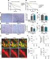
Fig. 5. Nec-1f protects from calcium oxalate (CaOx)-induced nephropathy and reduces neutrophil recruitment after heart transplantation.
A Human microsomal stability for Nec-1f in comparison with reference substances diclofenac and propanolol. B Human plasma stability of Nec-1f was assessed in comparison with reference substances (propantheline, verapamil) over 120 min. C Mice received a single injection of 100 mg/kg sodium oxalate and were supplemented with 3% sodium oxalate in the drinking water upon treatment with the vehicle of 1.65 mg/kg Nec-1f. Pizzolato staining visualizes calcium oxalate crystal deposition in the kidneys. Quantification of CaOx crystal deposition demonstrated equal amounts of deposition in each group. D Periodic acid-Schiff (PAS) staining in kidneys after CaOx treatment was used to assess the tubular damage. E Serum urea and F serum creatinine levels were analyzed 24 h after the injection of CaOx. The bar graphs (CF) show mean +/− SD. Statistical analysis was performed using two-sided students t-test. G Two-photon intravital imaging of neutrophil (green) behavior after transplantation of B6 hearts into control untreated (top) or Nec-1f-treated (2 mg/kg) (bottom) B6 LysM-GFP mice at indicated time points after reperfusion. Vessels were labeled red after the injection of quantum dots (q-dots). n = 4 per experimental group. Scale bars: 30 μm. H Intravascular rolling velocities of neutrophils, I neutrophil recruitment per minute to coronary veins, J density of neutrophils, and K percentage of extravasated neutrophils in control cardiac grafts and after treatment of recipient mice with Nec-1f. HK Shows the mean +/− SEM. *p < 0.05; n.s. not significant. Statistics were performed using the Mann−Whitney U test.
Fig. 6
Fig. 6. Nec-1f protects against bilateral renal ischemia-reperfusion injury in mice.
C57Bl/6 N mice underwent bilateral ischemia as detailed in the methods section. A Representative images of 4-Hydroxynonenal (4HNE)-staining, quantified in (B, n = 4). C Representative periodic acid-Schiff (PAS)-stained histological sections are presented and quantified using the tubular damage score (D, n = 4). Serum concentrations of E creatinine (sham n = 2, 48 h IRI vehicle n = 12, Nec-1f n = 10, 72 h IRI vehicle n = 8, Nec-1f n = 6) and F urea (sham n = 2, 48 h IRI vehicle n = 12, Nec-1f n = 11, 72 h IRI vehicle n = 6, Nec-1f n = 6) were measured in two independent experiments 48 and 72 h following the onset of reperfusion. G F4/80 positivity was stained by immunohistochemistry and quantified by experienced nephropathologists (H, I, n = 5). Bar graphs represent the mean +/− SD. J Kaplan−Meier survival curves following severe renal ischemia-reperfusion injury upon pre-treatment with 2 mg Fer-1/kg body weight, 2 mg Nec-1s/kg body weight, 2 mg Nec-1f/kg body weight, or vehicle control are shown. Differences in the survival curves of vehicle compared to Fer-1 or Nec-1s are not statistically significant. All experiments were performed in a strictly double-blinded manner. Statistical analysis was performed using a two-sided student’s t-test. *p < 0.05; n.s. not significant.

References

    1. Huang, M. et al. Integrin-linked kinase deficiency in collecting duct principal cell promotes necroptosis of principal cell and contributes to kidney inflammation and fibrosis. J. Am. Soc. Nephrol.: JASN10.1681/asn.2018111162 (2019). - PMC - PubMed
    1. Liu W, et al. RGMb protects against acute kidney injury by inhibiting tubular cell necroptosis via an MLKL-dependent mechanism. Proc. Natl Acad. Sci. USA. 2018;115:E1475–e1484. doi: 10.1073/pnas.1716959115. - DOI - PMC - PubMed
    1. Martin-Sanchez, D. et al. TWEAK and RIPK1 mediate a second wave of cell death during AKI. Proc. Natl Acad. Sci. USA10.1073/pnas.1716578115 (2018). - PMC - PubMed
    1. Schreiber A, et al. Necroptosis controls NET generation and mediates complement activation, endothelial damage, and autoimmune vasculitis. Proc. Natl Acad. Sci. USA. 2017;114:E9618–e9625. doi: 10.1073/pnas.1708247114. - DOI - PMC - PubMed
    1. Xu, Y. et al. A role for tubular necroptosis in cisplatin-induced AKI. J. Am. Soc. Nephrol. 26, 2647–2658 (2015). - PMC - PubMed

Publication types

MeSH terms