. 2021 Jul 20;11(1):14819.
doi: 10.1038/s41598-021-94192-7.

Visual instrumental learning in blindsight monkeys

Affiliations

Visual instrumental learning in blindsight monkeys

Rikako Kato et al. Sci Rep. .

Abstract

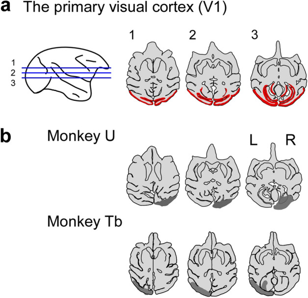
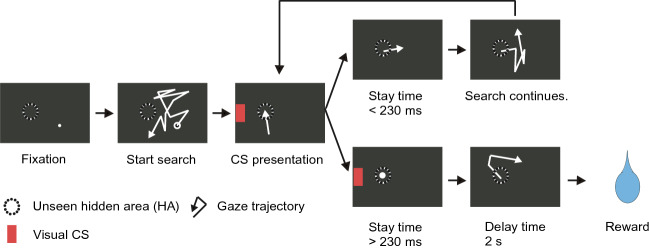
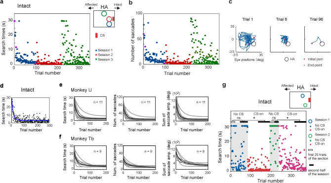
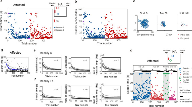
Blindsight is the residual visuo-motor ability without subjective awareness observed after lesions of the primary visual cortex (V1). Various visual functions are retained, however, instrumental visual associative learning remains to be investigated. Here we examined the secondary reinforcing properties of visual cues presented to the hemianopic field of macaque monkeys with unilateral V1 lesions. Our aim was to test the potential role of visual pathways bypassing V1 in reinforcing visual instrumental learning. When learning the location of a hidden area in an oculomotor search task, conditioned visual cues presented to the lesion-affected hemifield operated as an effective secondary reinforcer. We noted that not only the hidden area location, but also the vector of the saccade entering the target area was reinforced. Importantly, when the visual reinforcement signal was presented in the lesion-affected field, the monkeys continued searching, as opposed to stopping when the cue was presented in the intact field. This suggests the monkeys were less confident that the target location had been discovered when the reinforcement cue was presented in the affected field. These results indicate that the visual signals mediated by the residual visual pathways after V1 lesions can access fundamental reinforcement mechanisms but with impaired visual awareness.


Conflict of interest statement

The authors declare no competing interests.

Figures

Figure 1
Figure 1
Unilateral V1 lesion. Extent of unilateral V1 lesion in monkeys U, Tb. (a) and (b) MR images of horizontal sections of the monkey’s brain were traced and the extent of lesion was drawn on the traces. (a) The bilateral V1 is shown in red on the trace of monkey U before lesion. (b) The lesion sites (gray area) of monkeys U and Tb on the traces. Lesion sites for monkey U and monkey Tb were described in previous papers,. However, the horizontal sections of monkey U and monkey Tb in (b) were obtained from more recent MR images compared to those published in the previous literature.
Figure 2
Figure 2
Hidden target area search task. Sequence of trial events in the search task. In each trial the offset of randomly located fixation point signaled that the search for the unseen hidden area (HA) could begin. When the monkey’s gaze entered the HA, a reinforcing visual CS was presented.
Figure 3
Figure 3
Instrumental learning with CS in the intact visual field. (a) and (b) Examples of reduction in search time (a) and number of saccades (b) when the CS was presented in intact field. Search time and number of saccades for each trial was plotted against the trial number. HA position was altered in each session. (c) Eye position trajectories for three trials (trial 1, trial 8, trial 96) of session 1. The three trials are labeled by magenta circle in (a) and (b). (d) The optimally fitting decaying exponential curves (blue line) are superimposed on an example dataset for which the search time are plotted against the trial number in the session. The CS was presented in the intact visual hemifield. (e) and (f) Population data that characterize the learning of HA location in the instrumental conditioning task. (e) Population data from monkey U. Decaying exponential fitting curves from sessions in the intact condition were superimposed. Means of the curves are indicated as thick black lines. (f) The population data from monkey Tb, the same arrangement as (e). (g) No CS condition. Learning failed to occur in the No CS conditions (grey hatched area) but was reinstated when the CS was presented to the intact field. *Wilcoxon rank sum test, p < 0.05.
Figure 4
Figure 4
Instrumental learning with CS in the affected visual field. (a) and (b) Examples of reduction in search time (a) and number of saccades (b) when the CS was presented in the affected field. Search time and number of saccades for each trial was plotted against the trial number. HA position was altered in each session. (c) Eye position trajectories for three trials (trial 3, trial 69, trial 176) of session 1. The three trials are labeled by magenta circle in (a) and (b). (d) The optimally fitting decaying exponential curves (blue line) are superimposed on an example dataset for which the search time are plotted against the trial number in the session. (e) and (f) Population data that characterize the learning of HA location in the instrumental conditioning task. (e) Population data from monkey U. Decaying exponential fitting curves from sessions in the affected condition were superimposed. Means of the curves are indicated as thick black lines. (f) The population data from monkey Tb, the same arrangement as (e). (g) No CS condition. Learning failed to occur in the No CS conditions (grey hatched area) but was reinstated when the CS was presented to the affected field. *Wilcoxon rank sum test, p < 0.05.
Figure 5
Figure 5
Learning the matrix of the final saccades that preceded CS presentation. (a) Schematic illustration of a final saccade capturing the HA and its parameters. (b) and (c) Vectors of final saccades when the CS was presented in the lesion-affected field. The final saccades of the 20 successful trials among the trial No. 1–34 (b) and trial No. 80–99 (c) of a session which is the first session in Fig. 4a, b. A final saccade was characterized by three parameters [start point (d), direction (e) and amplitude (f)] of the vector from its onset location to its offset in the HA (a). (d)–(f) Saccade parameters in a session, shown in (b) and (c). Start point (d), direction (e) and amplitude (f) of the final saccades. (g) and (h) Distribution of variance of direction of the final saccades in multiple sessions. (g) CS in the intact field and (h) lesion-affected field.
Figure 6
Figure 6
Patterns of eye movements differed after CS presentation to the intact and lesion-affect visual fields. (a)–(d) During a single search trial, saccades before (blue lines) and after (red lines) CS presentation to the intact (a) and lesion-affected (b) visual fields. In a whole session, the distributions of saccade end points (ep) before (blue) and after (red) CS presentation to the intact (c) and lesion-affect (d) visual fields. (e) Spatial homogeneity of distributions of saccade end points in each session calculated by modified Rippley’s K function between before and after CS presentation to the intact and lesion-affected visual fields (mK).

Similar articles

Cited by

References

    1. Pöppel E, Held R, Frost D. Residual visual function after Brain Wounds involving the central visual pathways in Man. Nature. 1973;243:295–296. doi: 10.1038/243295a0. - DOI - PubMed
    1. Sanders MD, Warrington EK, Marshall J, Wieskrantz L. ‘BLINDSIGHT’: Vision in a field defect. Lancet. 1974;303:707–708. doi: 10.1016/S0140-6736(74)92907-9. - DOI - PubMed
    1. Weiskrantz L, Warrington E, K., Sanders, M. D. & Marshall, J. Visual capacity in the hemianopic field following a restricted occipital ablation. Brain. 1974;97:709–728. doi: 10.1093/brain/97.1.709. - DOI - PubMed
    1. Cowey A. The blindsight saga. Exp. Brain Res. 2010;200:3–24. doi: 10.1007/s00221-009-1914-2. - DOI - PubMed
    1. Tamietto M, Morrone MC. Visual plasticity: Blindsight bridges anatomy and function in the visual system. Curr. Biol. 2016;26(2):70–73. doi: 10.1016/j.cub.2015.11.026. - DOI - PMC - PubMed

Publication types