

Efficient gene transfer into zebra finch germline-competent stem cells using an adenoviral vector system
- PMID: 34285320
- PMCID: PMC8292312
- DOI: 10.1038/s41598-021-94229-x
Efficient gene transfer into zebra finch germline-competent stem cells using an adenoviral vector system
Abstract
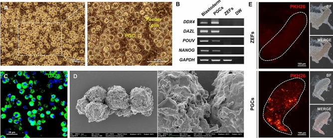
Zebra finch is a representative animal model for studying the molecular basis of human disorders of vocal development and communication. Accordingly, various functional studies of zebra finch have knocked down or introduced foreign genes in vivo; however, their germline transmission efficiency is remarkably low. The primordial germ cell (PGC)-mediated method is preferred for avian transgenic studies; however, use of this method is restricted in zebra finch due to the lack of an efficient gene transfer method for the germline. To target primary germ cells that are difficult to transfect and manipulate, an adenovirus-mediated gene transfer system with high efficiency in a wide range of cell types may be useful. Here, we isolated and characterized two types of primary germline-competent stem cells, PGCs and spermatogonial stem cells (SSCs), from embryonic and adult reproductive tissues of zebra finch and demonstrated that genes were most efficiently transferred into these cells using an adenovirus-mediated system. This system was successfully used to generate gene-edited PGCs in vitro. These results are expected to improve transgenic zebra finch production.
© 2021. The Author(s).
Conflict of interest statement
The authors declare no competing interests.
Figures


Similar articles
-
Identification and characterization of primordial germ cells in a vocal learning Neoaves species, the zebra finch.FASEB J. 2019 Dec;33(12):13825-13836. doi: 10.1096/fj.201900760RR. Epub 2019 Oct 11. FASEB J. 2019. PMID: 31604057
-
Pronounced early differentiation underlies zebra finch gonadal germ cell development.Dev Biol. 2025 Jan;517:73-90. doi: 10.1016/j.ydbio.2024.08.006. Epub 2024 Aug 28. Dev Biol. 2025. PMID: 39214328
-
Highly Efficient Genome Modification of Cultured Primordial Germ Cells with Lentiviral Vectors to Generate Transgenic Songbirds.Stem Cell Reports. 2021 Apr 13;16(4):784-796. doi: 10.1016/j.stemcr.2021.02.015. Epub 2021 Mar 18. Stem Cell Reports. 2021. PMID: 33740464 Free PMC article.
-
Current Approaches and Applications in Avian Genome Editing.Int J Mol Sci. 2020 May 30;21(11):3937. doi: 10.3390/ijms21113937. Int J Mol Sci. 2020. PMID: 32486292 Free PMC article. Review.
-
Gene transfer strategies for the physiologist.Exp Physiol. 2000 Nov;85(6):735-45. doi: 10.1111/j.1469-445x.2000.02142.x. Exp Physiol. 2000. PMID: 11187967 Review.
Cited by
-
Generation and characterization of genome-modified chondrocyte-like cells from the zebra finch cell line immortalized by c-MYC expression.Front Zool. 2022 Jun 11;19(1):18. doi: 10.1186/s12983-022-00464-x. Front Zool. 2022. PMID: 35690812 Free PMC article.
-
Generation of genome-edited chicken and duck lines by adenovirus-mediated in vivo genome editing.Proc Natl Acad Sci U S A. 2022 Nov 8;119(45):e2214344119. doi: 10.1073/pnas.2214344119. Epub 2022 Nov 2. Proc Natl Acad Sci U S A. 2022. PMID: 36322747 Free PMC article.
-
Induction of an immortalized songbird cell line allows for gene characterization and knockout by CRISPR-Cas9.Sci Rep. 2022 Mar 14;12(1):4369. doi: 10.1038/s41598-022-07434-7. Sci Rep. 2022. PMID: 35288582 Free PMC article.
-
A Model of Discovery: The Role of Imaging Established and Emerging Non-mammalian Models in Neuroscience.Front Mol Neurosci. 2022 Apr 14;15:867010. doi: 10.3389/fnmol.2022.867010. eCollection 2022. Front Mol Neurosci. 2022. PMID: 35493325 Free PMC article. Review.
-
Propagation of goose primordial germ cells in vitro relies on FGF and BMP signalling pathways.Commun Biol. 2025 Feb 25;8(1):301. doi: 10.1038/s42003-025-07715-7. Commun Biol. 2025. PMID: 40000797 Free PMC article.
References
Publication types
MeSH terms
Substances
LinkOut - more resources
Full Text Sources
