Review
. 2021 Jul 5:2021:6670579.
doi: 10.1155/2021/6670579. eCollection 2021.

The Role of Mitochondria in Liver Ischemia-Reperfusion Injury: From Aspects of Mitochondrial Oxidative Stress, Mitochondrial Fission, Mitochondrial Membrane Permeable Transport Pore Formation, Mitophagy, and Mitochondria-Related Protective Measures

Affiliations
Review

The Role of Mitochondria in Liver Ischemia-Reperfusion Injury: From Aspects of Mitochondrial Oxidative Stress, Mitochondrial Fission, Mitochondrial Membrane Permeable Transport Pore Formation, Mitophagy, and Mitochondria-Related Protective Measures

Haifeng Zhang et al. Oxid Med Cell Longev. .

Abstract

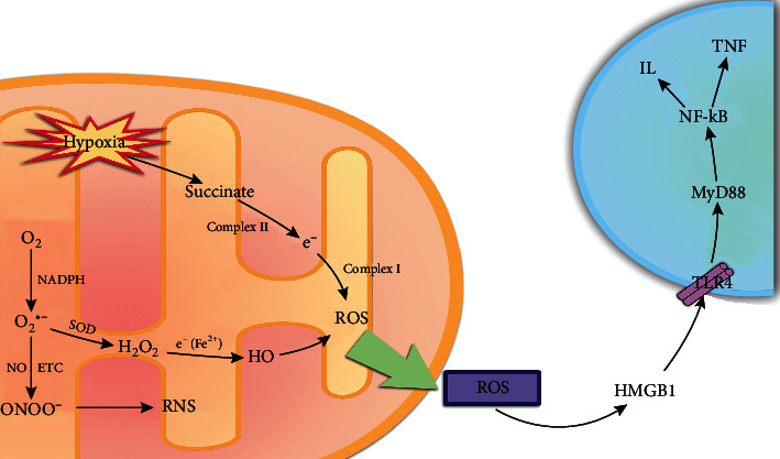
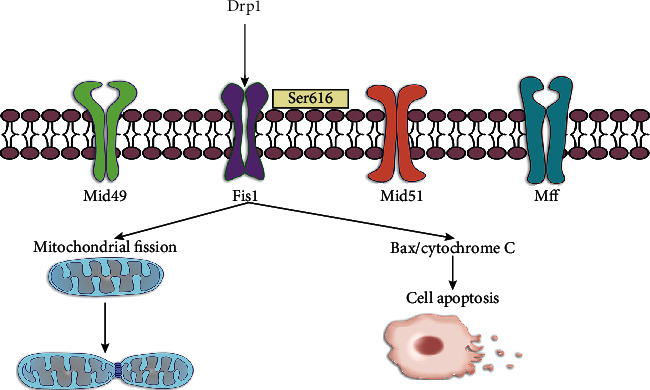
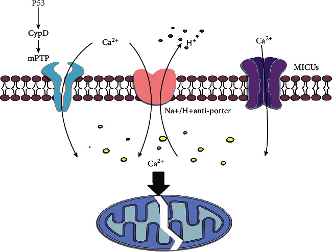
Ischemia-reperfusion injury (IRI) has indeed been shown as a main complication of hepatectomy, liver transplantation, trauma, and hypovolemic shock. A large number of studies have confirmed that microvascular and parenchymal damage is mainly caused by reactive oxygen species (ROS), which is considered to be a major risk factor for IRI. Under normal conditions, ROS as a kind of by-product of cellular metabolism can be controlled at normal levels. However, when IRI occurs, mitochondrial oxidative phosphorylation is inhibited. In addition, oxidative respiratory chain damage leads to massive consumption of adenosine triphosphate (ATP) and large amounts of ROS. Additionally, mitochondrial dysfunction is involved in various organs and tissues in IRI. On the one hand, excessive free radicals induce mitochondrial damage, for instance, mitochondrial structure, number, function, and energy metabolism. On the other hand, the disorder of mitochondrial fusion and fission results in further reduction of the number of mitochondria so that it is not enough to clear excessive ROS, and mitochondrial structure changes to form mitochondrial membrane permeable transport pores (mPTPs), which leads to cell necrosis and apoptosis, organ failure, and metabolic dysfunction, increasing morbidity and mortality. According to the formation mechanism of IRI, various substances have been discovered or synthesized for specific targets and cell signaling pathways to inhibit or slow the damage of liver IRI to the body. Here, based on the development of this field, this review describes the role of mitochondria in liver IRI, from aspects of mitochondrial oxidative stress, mitochondrial fusion and fission, mPTP formation, and corresponding protective measures. Therefore, it may provide references for future clinical treatment and research.


Conflict of interest statement

The authors declare no conflicts of interest that pertain to this work.

Figures

Figure 1
Figure 1
The arrows refer to the role of promotion, and the symbol of “┴” refers to the role of inhibition. Under the action of NADPH, O2 can produce O2•−, which is considered to be one of the most important ROS. On the one hand, O2•− can react with super NO to produce ONOO. On the other hand, two molecules of O2•− can generate H2O2 and one molecule of water under the action of SOD. The H2O2 obtains electrons from ferrous ions to generate HO. In addition, hypoxia leads to the accumulation of succinate and its metabolites. A large amount of accumulated succinate is oxidized by complex II and further transmits electrons to complex I and generates a large amount of ROS. Further, ROS passes through HMGB1-TLR1-MyD88-NF-κB signaling pathway stimulates the release of inflammatory factors and induces cell apoptosis.
Figure 2
Figure 2
The arrows refer to the role of promotion, and the symbol of “┴” refers to the role of inhibition. Under normal circumstances, mitochondria contain the four receptors of Drp1 (Mid49, Mid51, Fis1, and Mff). The phosphorylation of Drp1 at Ser-616 causes mitochondrial fission and hepatocyte apoptosis.
Figure 3
Figure 3
The arrows refer to the role of promotion, and the symbol of “┴” refers to the role of inhibition. Na+/H+ antiporter and mitochondrial Ca2+ uptake 1/2 (MICUs), and the P53-CypD signaling pathway is an important regulatory pathway for mPTP formation and calcium overload. Excessive calcium concentration in mitochondria can induce mitochondrial damage and apoptosis.

References

    1. Takanori K., Lentsch A. B. Hepatic ischemia/reperfusion: mechanisms of tissue injury, repair, and regeneration. Gene Expression. 2017;17(4):277–287. doi: 10.3727/105221617x15042750874156. - DOI - PMC - PubMed
    1. Ito T., Naini B. V., Markovic D., et al. Ischemia-reperfusion injury and its relationship with early allograft dysfunction in liver transplant patients. American Journal of Transplantation. 2021;21(2):614–625. doi: 10.1111/ajt.16219. - DOI - PubMed
    1. Huang M., du J., Wang Y., et al. Tissue kallikrein-related peptidase8 protects rat heart against acute ischemia reperfusion injury. International Journal of Biological Macromolecules. 2019;140:1126–1133. doi: 10.1016/j.ijbiomac.2019.08.195. - DOI - PubMed
    1. Klevstig M., Arif M., Mannila M., et al. Cardiac expression of the microsomal triglyceride transport protein protects the heart function during ischemia. Journal of Molecular and Cellular Cardiology. 2019;137:1–8. doi: 10.1016/j.yjmcc.2019.09.003. - DOI - PubMed
    1. Aguilar A. Loss of PKC-ɛ protects against IRI. Nephrology. 2016;12(12):p. 714. doi: 10.1038/nrneph.2016.162. - DOI - PubMed

MeSH terms

Substances

LinkOut - more resources