

Curculigoside Protects against Titanium Particle-Induced Osteolysis through the Enhancement of Osteoblast Differentiation and Reduction of Osteoclast Formation
- PMID: 34285923
- PMCID: PMC8275416
- DOI: 10.1155/2021/5707242
Curculigoside Protects against Titanium Particle-Induced Osteolysis through the Enhancement of Osteoblast Differentiation and Reduction of Osteoclast Formation
Abstract
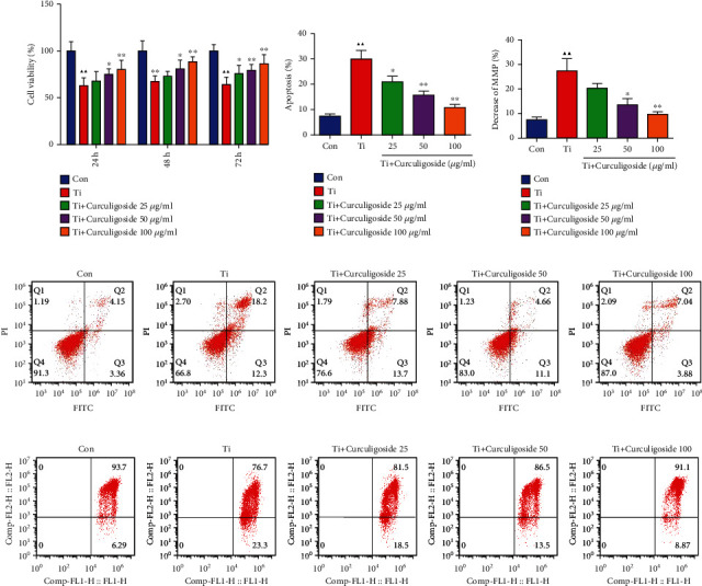
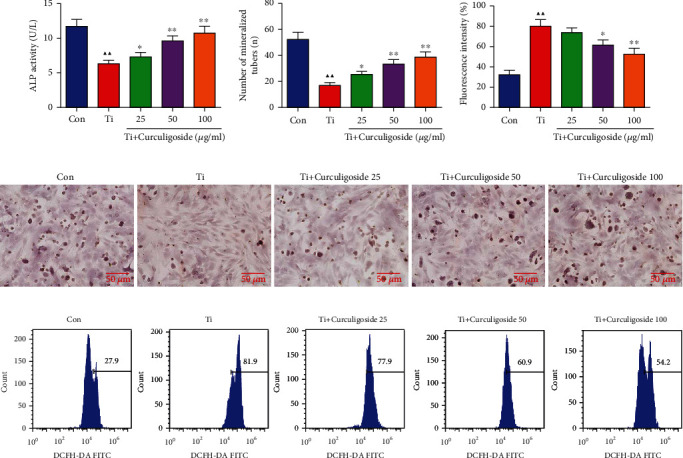
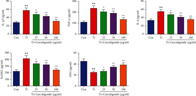
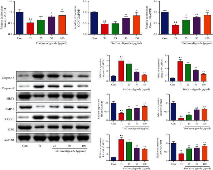
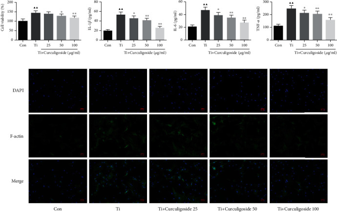
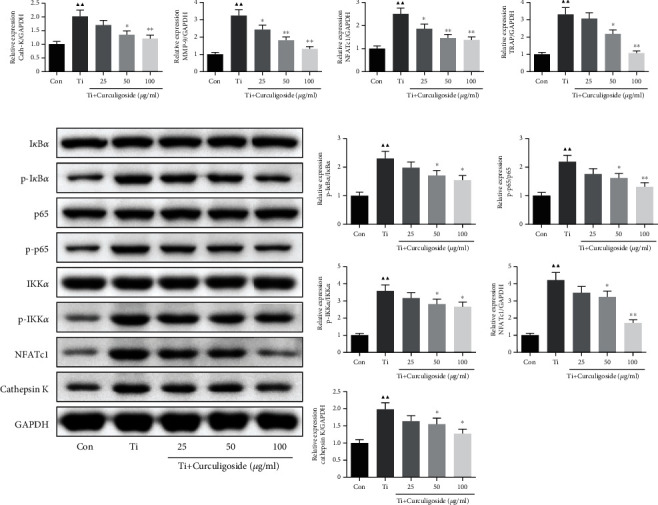
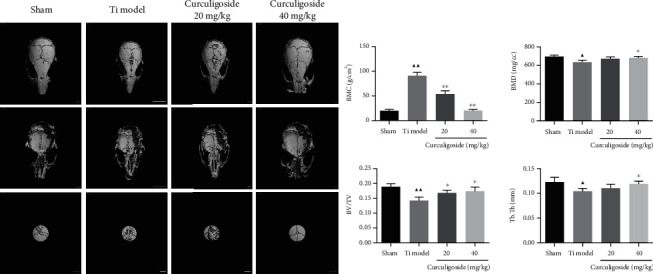
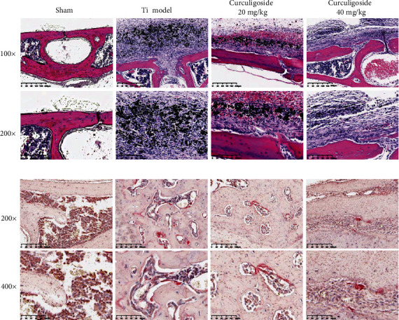
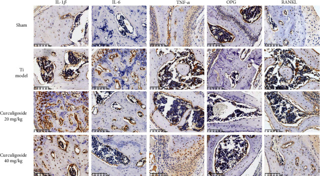
Wear particle-induced periprosthetic osteolysis is mainly responsible for joint replacement failure and revision surgery. Curculigoside is reported to have bone-protective potential, but whether curculigoside attenuates wear particle-induced osteolysis remains unclear. In this study, titanium particles (Ti) were used to stimulate osteoblastic MC3T3-E1 cells in the presence or absence of curculigoside, to determine their effect on osteoblast differentiation. Rat osteoclastic bone marrow stromal cells (BMSCs) were cocultured with Ti in the presence or absence of curculigoside, to evaluate its effect on osteoclast formation in vitro. Ti was also used to stimulate mouse calvaria to induce an osteolysis model, and curculigoside was administrated to evaluate its effect in the osteolysis model by micro-CT imaging and histopathological analyses. As the results indicated, in MC3T3-E1 cells, curculigoside treatment attenuated the Ti-induced inhibition on cell differentiation and apoptosis, increased alkaline phosphatase activity (ALP) and cell mineralization, and inhibited TNF-α, IL-1β, and IL-6 production and ROS generation. In BMSCs, curculigoside treatment suppressed the Ti-induced cell formation and suppressed the TNF-α, IL-1β, and IL-6 production and F-actin ring formation. In vivo, curculigoside attenuated Ti-induced bone loss and histological damage in murine calvaria. Curculigoside treatment also reversed the RANK/RANKL/OPG and NF-κB signaling pathways, by suppressing the RANKL and NF-κB expression, while activating the OPG expression. Our study demonstrated that curculigoside treatment was able to attenuate wear particle-induced periprosthetic osteolysis in in vivo and in vitro experiments, promoted osteoblastic MC3T3-E1 cell differentiation, and inhibited osteoclast BMSC formation. It suggests that curculigoside may be a potential pharmaceutical agent for wear particle-stimulated osteolysis therapy.
Copyright © 2021 Fangbing Zhu et al.
Conflict of interest statement
The authors declare that there are no conflicts of interest.
Figures
Similar articles
-
Curculigoside is a Promising Osteoprotective Agent for Osteoporosis: Review.Drug Des Devel Ther. 2025 Apr 28;19:3323-3336. doi: 10.2147/DDDT.S519174. eCollection 2025. Drug Des Devel Ther. 2025. PMID: 40322028 Free PMC article. Review.
-
Pentamidine Inhibits Titanium Particle-Induced Osteolysis In Vivo and Receptor Activator of Nuclear Factor-κB Ligand-Mediated Osteoclast Differentiation In Vitro.Tissue Eng Regen Med. 2019 Apr 2;16(3):265-273. doi: 10.1007/s13770-019-00186-y. eCollection 2019 Jun. Tissue Eng Regen Med. 2019. PMID: 31205855 Free PMC article.
-
Titanium particle-challenged osteoblasts promote osteoclastogenesis and osteolysis in a murine model of periprosthestic osteolysis.Acta Biomater. 2013 Jul;9(7):7564-72. doi: 10.1016/j.actbio.2013.03.010. Epub 2013 Mar 19. Acta Biomater. 2013. PMID: 23518478 Free PMC article.
-
Inhibitory effects of triptolide on titanium particle-induced osteolysis and receptor activator of nuclear factor-κB ligand-mediated osteoclast differentiation.Int Orthop. 2015 Jan;39(1):173-82. doi: 10.1007/s00264-014-2596-3. Epub 2014 Nov 23. Int Orthop. 2015. PMID: 25416122
-
The dual role of autophagy in periprosthetic osteolysis.Front Cell Dev Biol. 2023 Mar 24;11:1123753. doi: 10.3389/fcell.2023.1123753. eCollection 2023. Front Cell Dev Biol. 2023. PMID: 37035243 Free PMC article. Review.
Cited by
-
Epimedium-Curculigo herb pair enhances bone repair with infected bone defects and regulates osteoblasts through LncRNA MALAT1/miR-34a-5p/SMAD2 axis.J Cell Mol Med. 2024 Jul;28(13):e18527. doi: 10.1111/jcmm.18527. J Cell Mol Med. 2024. PMID: 38984969 Free PMC article.
-
The role of mitochondria in the peri-implant microenvironment.Exp Physiol. 2023 Mar;108(3):398-411. doi: 10.1113/EP090988. Epub 2023 Jan 17. Exp Physiol. 2023. PMID: 36648334 Free PMC article. Review.
-
Titanium particles inhibit bone marrow mesenchymal stem cell osteogenic differentiation through the MAPK signaling pathway.FEBS Open Bio. 2023 Sep;13(9):1699-1708. doi: 10.1002/2211-5463.13678. Epub 2023 Jul 30. FEBS Open Bio. 2023. PMID: 37483149 Free PMC article.
-
Curculigoside is a Promising Osteoprotective Agent for Osteoporosis: Review.Drug Des Devel Ther. 2025 Apr 28;19:3323-3336. doi: 10.2147/DDDT.S519174. eCollection 2025. Drug Des Devel Ther. 2025. PMID: 40322028 Free PMC article. Review.
-
Human umbilical cord mesenchymal stromal cells promotes the proliferation and osteogenic differentiation of autologous bone marrow stem cells by secreting exosomes.Bioengineered. 2022 Apr;13(4):9901-9915. doi: 10.1080/21655979.2022.2062183. Bioengineered. 2022. PMID: 35412945 Free PMC article.
References
-
- Evans J. T., Evans J. P., Walker R. W., Blom A. W., Whitehouse M. R., Sayers A. How long does a knee replacement last? A systematic review and meta-analysis of case series and national registry reports with more than 15 years of follow-up. The Lancet. 2019;393(10172):655–663. doi: 10.1016/S0140-6736(18)32531-5. - DOI - PMC - PubMed
-
- Evans J. T., Evans J. P., Walker R. W., Blom A. W., Whitehouse M. R., Sayers A. How long does a hip replacement last? A systematic review and meta-analysis of case series and national registry reports with more than 15 years of follow-up. The Lancet. 2019;393(10172):647–654. doi: 10.1016/S0140-6736(18)31665-9. - DOI - PMC - PubMed
MeSH terms
Substances
LinkOut - more resources
Full Text Sources
Research Materials
