

3D Printing of Layered Gradient Pore Structure of Brain-like Tissue
- PMID: 34286148
- PMCID: PMC8287709
- DOI: 10.18063/ijb.v7i3.359
3D Printing of Layered Gradient Pore Structure of Brain-like Tissue
Abstract
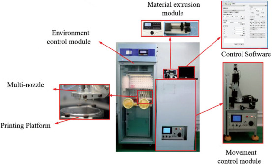
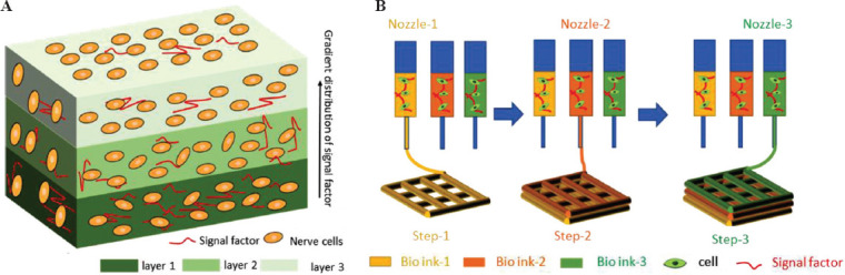
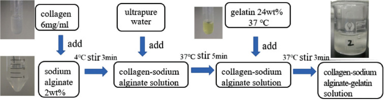
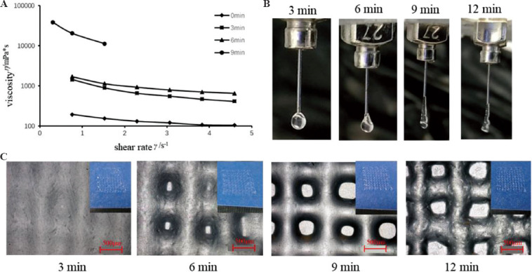
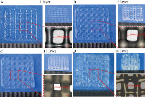
The pathological research and drug development of brain diseases require appropriate brain models. Given the complex, layered structure of the cerebral cortex, as well as the constraints on the medical ethics and the inaccuracy of animal models, it is necessary to construct a brain-like model in vitro. In this study, we designed and built integrated three-dimensional (3D) printing equipment for cell printing/culture, which can guarantee cell viability in the printing process and provide the equipment foundation for manufacturing the layered structures with gradient distribution of pore size. Based on this printing equipment, to achieve the purpose of printing the layered structures with multiple materials, we conducted research on the performance of bio-inks with different compositions and optimized the printing process. By extruding and stacking materials, we can print the layered structure with the uniform distribution of cells and the gradient distribution of pore sizes. Finally, we can accurately print a structure with 30 layers. The line width (resolution) of the printed monolayer structure was about 478 mm, the forming accuracy can reach 97.24%, and the viability of cells in the printed structure is as high as 94.5%.
Keywords: 3D bio-printing; Brain-like model; Integrated cell printing/culture equipment; Layered gradient structure.
Copyright: © 2021 Pei, et al.
Conflict of interest statement
We have no financial and personal relationships with other people or organizations that can inappropriately influence our work, there is no professional or other personal interest of any nature or kind in any product, service, and/or company that could be construed as influencing the position presented in, or the review of, this article.
Figures

References
-
- Muming Pu BX. Nao Ke Xue Yu Lei Nao Yan Jiu Gai Shu. [Brain Science and Brain-Inspired Intelligence Technology] J Chin Acad Sci. 2016;31:725–36.
-
- Xiong Y, Mahmood A, Chopp M. Animal Models of Traumatic Brain Injury. Nat Rev Neurosci. 2013;14:128–42. https://doi.org/10.1038/nrn3407. - PMC - PubMed
-
- Huh D, Hamilton GA, Ingber DE. From 3D Cell Culture to Organs-on-Chips. Trends Cell Biol. 2011;21:745–54. https://doi.org/10.1016/j.tcb.2011.09.005. - PMC - PubMed
-
- Imamura Y, Mukohara T, Shimono Y, et al. Comparison of 2D- and 3D-Culture Models as Drug-testing Platforms in Breast Cancer. Oncol Rep. 2015;33:1837–43. https://doi.org/10.3892/or.2015.3767. - PubMed
-
- Tian XF, Heng BC, Ge Z, et al. Comparison of Osteogenesis of Human Embryonic Stem Cells within 2D and 3D Culture systems. Scand J Clin Lab Invest. 2008;68:58–6. https://doi.org/10.1080/00365510701466416. - PubMed
LinkOut - more resources
Full Text Sources
Other Literature Sources