Review
. 2021 Jul 9;5(3):031503.
doi: 10.1063/5.0046682. eCollection 2021 Sep.

Functional hydrogels for diabetic wound management

Affiliations
Review

Functional hydrogels for diabetic wound management

Daqian Gao et al. APL Bioeng. .

Abstract

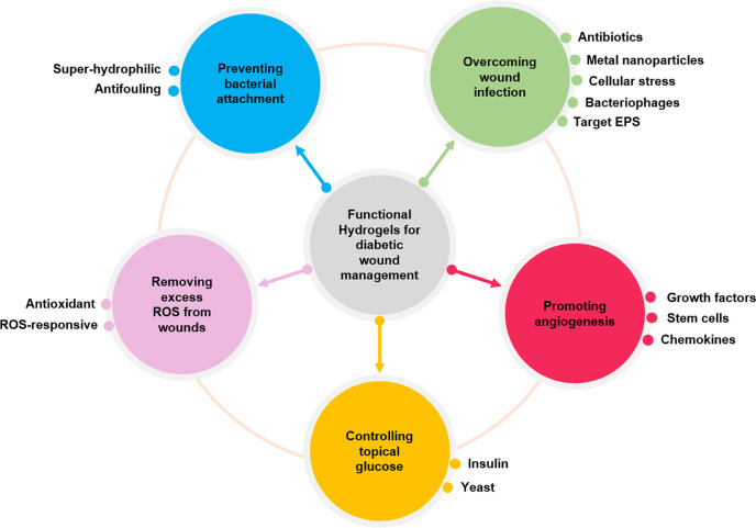
Diabetic wounds often have a slow healing process and become easily infected owing to hyperglycemia in wound beds. Once planktonic bacterial cells develop into biofilms, the diabetic wound becomes more resistant to treatment. Although it remains challenging to accelerate healing in a diabetic wound due to complex pathology, including bacterial infection, high reactive oxygen species, chronic inflammation, and impaired angiogenesis, the development of multifunctional hydrogels is a promising strategy. Multiple functions, including antibacterial, pro-angiogenesis, and overall pro-healing, are high priorities. Here, design strategies, mechanisms of action, performance, and application of functional hydrogels are systematically discussed. The unique properties of hydrogels, including bactericidal and wound healing promotive effects, are reviewed. Considering the clinical need, stimuli-responsive and multifunctional hydrogels that can accelerate diabetic wound healing are likely to form an important part of future diabetic wound management.


Figures

FIG. 1.
FIG. 1.
Functional hydrogels for diabetic wound management.
FIG. 2.
FIG. 2.
Zwitterionic hydrogels for preventing bacterial attachment: (a) a zwitterionic polyampholyte hydrogel was grafted on a polyethersulfone (PES) surface to provide antifouling properties. Reprinted with permission from Wang et al., J. Membr. Sci. 565, 293–302 (2018). Copyright 2018 Elsevier. (b) A zwitterionic hydrogel crosslinked via the thiol–ene click reaction resisted adhesion of protein and cells. Reproduced with permission from Guo et al., Chem. Mater. 32(15), 6347–6357 (2020). Copyright 2020 American Chemical Society.
FIG. 3.
FIG. 3.
In vitro eradication of established biofilms using hydrogels loaded with antibacterial components: (a) different concentrations of ultrasmall silver nanoparticles (AgNPs) (25 and 50 μg/g gel) were incorporated into Pluronic F-127 hydrogels to destroy the structures of P. aeruginosa biofilms. Reproduced with permission from Haidari et al., ACS Appl. Mater. Interfaces 12(37), 41011–41025 (2020). Copyright 2020 American Chemical Society. (b) Dextran methacrylate hydrogel was synthesized by photopolymerization polymer for encapsulating antibacterial cationic small molecules, which can penetrate the extracellular polymeric substances (EPS) of the established S. aureus and E. coli biofilms. Reproduced with permission from Hoque et al., ACS Appl. Mater. Interfaces 9(19), 15975–15985 (2017). Copyright 2017 American Chemical Society.
FIG. 4.
FIG. 4.
Synergy between antifouling and antibacterial hydrogels: (a) a zwitterionic hydrogel was obtained by simply adjusting the ratio between cationic chitosan and anionic dextran while incorporating AgNPs for enhancing their antibacterial activity, (b) the hydrogels with AgNPs showed superior antibacterial properties. Reproduced with permission from Shi et al., Langmuir 35(5), 1837–1845 (2019). Copyright 2018 American Chemical Society. (c) A hybrid hydrogel nanoneedle surface that can rupture cell membranes was fabricated based on zwitterionic polymers via a UV replica molding technique, providing antifouling and antibacterial properties, (d) E. coli was cultured on the 2-methacryloyloxyethyl phosphorylcholine (MPC)-grafted planar surface and MPC-grafted nanoneedle surfaces prepared with polyurethane acrylate (PUA) and different concentrations (80%, 90%, and 100%) of poly(ethylene glycol) dimethacrylate (PEGDMA). MPC-grafted nanoneedle surfaces prevented biofilm formation. Reproduced with permission from Park et al., ACS Macro Lett. 8(1), 64–69 (2019). Copyright 2018 American Chemical Society.
FIG. 5.
FIG. 5.
Smart hydrogels for antifouling: (a) using a salt-responsive surface based on a polyzwitterionic polymer, the contact angle decreased when the solution changed from water to 1 M NaCl solution; thus, bacteria were killed in water and released in salt water. Reprinted with permission from Huang et al., Chem. Eng. J. 333, 1–10 (2017). Copyright 2017Elsevier. (b) The surface of hydrogel based on photoresponsive polymer, poly[2-((4,5-dimethoxy-2-nitrobenzyl)oxy)-N-(2-(methacryloyloxy)ethyl)-N,N-dimethyl-2-oxoethan-1-aminium] (polyCBNA), was switched from cationic antibacterial to zwitterionic antifouling by UV treatment. Reproduced with permission from Liu et al., Langmuir 35(5), 1450–1457 (2019). Copyright 2018 American Chemical Society. (c) A thermosensitive poly(N-isopropylacrylamide) (PNIPAAm) hydrogel encapsulating AgNPs was prepared via a photopolymerization method. As temperature changed from 37 °C to 4 °C, the surface switched from killing to repelling bacteria mode. Reproduced with permission from Yang et al., ACS Appl. Mater. Interfaces 8(41), 28047–28054 (2016). Copyright 2016 American Chemical Society.
FIG. 6.
FIG. 6.
ROS-scavenging hydrogel: a polyvinyl alcohol (PVA) hydrogel was crosslinked by a ROS-responsive crosslinker (on the left). 100% of H2O2 was scavenged after 24 h, when the hydrogel was incubated in H2O2 (1 mM, 2 ml) solution (on the right). Reprinted with permission from Zhao et al., Biomaterials 258, 120286 (2020). Copyright 2020 Elsevier.
FIG. 7.
FIG. 7.
Hydrogel promoting angiogenesis: (a) the combination of desferrioxamine (DFO) and bioglass (BG) loaded sodium alginate (SA) hydrogels affected the migration and tube formation of HUVECs in vitro and (b) also promoted angiogenic factor expression in vivo (immunohistochemical staining for HIF-1α and VEGF). Reproduced with permission from Kong et al., ACS Appl. Mater. Interfaces 10(36), 30103–30114 (2018). Copyright 2018 American Chemical Society.
FIG. 8.
FIG. 8.
Multifunctional hydrogels for diabetic wounds with persistent infections: (a) a schematic diagram illustrating the injectable, self-healing, and adhesive hydrogel (FEMI) hydrogel for multidrug-resistant (MDR) bacteria-infected diabetic wound healing. After loading insulin and MnO2 nanosheets, the hydrogel can regulate blood glucose and relieve oxidative stress of wounds. Reproduced with permission from Wang et al., Nano Lett. 20(7), 5149–5158 (2020). Copyright 2020 American Chemical Society. (b) A preparation scheme of a yeast immobilized and copper nanoparticle incorporated wound dressing, the dressing can produce ethanol by consuming glucose, helping to control bacteria growth. Reproduced with permission from Bhadauriya et al., ACS Appl. Bio Mater. 1(2), 246–258 (2018). Copyright 2018 American Chemical Society.
FIG. 9.
FIG. 9.
Hydrogels for promoting angiogenesis and anti-infection: (a) self-healing Ag(I)-thiol (Au–S) coordinative hydrogel was developed by mixing four-arm-PEG-SH with AgNO3 that possessed antibacterial, pro-angiogenesis, and pro-epithelization properties. Reproduced with permission from Chen et al., NPG Asia Mater. 11, 3 (2019). Copyright 2019 Authors, licensed under a Creative Commons Attribution (CC BY) license. (b) Schematic illustration of multifunctional polysaccharide-based (FEP) hydrogel loaded with exosomes to accelerate diabetic wound healing. Reproduced with permission from Wang et al., ACS Nano 13(9), 10279–10293 (2019). Copyright 2019 American Chemical Society.
FIG. 10.
FIG. 10.
Smart hydrogel wound dressings that sense, monitor, and interact with wounds while treating chronic infections in diabetic wounds: (a) glucose-sensing enzymes that can react in proportion to glucose concentration and a pH dye were immobilized into an anti‐biofouling zwitterionic hydrogel, which provided a moist healing environment for diabetic wounds. Meanwhile, glucose concentration between 0 and 10 mM and pH value (4.0–8.0) in wound microenvironment can be monitored. Reproduced with permission from Zhu et al., Adv. Funct. Mater. 30(6), 1905493 (2020). Copyright 2019 Wiley‐VCH Verlag GmbH & Co. KGaA, Weinheim. (b) A thermosensitive chitosan (CS)-based hydrogel encapsulating pH-sensitive dye and near-infrared (NIR)-absorbing conjugated polymer can detect the pH change of an infected wound. Then photothermal therapy after in situ visual diagnosis. Reproduced with permission from Wang et al., ACS Appl. Mater. Interfaces 12(35), 39685–39694 (2020). Copyright 2020 American Chemical Society.
FIG. 11.
FIG. 11.
On-demand wound dressing: electrically driven responsive thread-based antibacterial wound patch with conductive, thermo-responsive, and drug-releasing properties. Reproduced with permission from Mostafalu et al., Adv. Funct. Mater. 27(41), 1702399 (2017). Copyright 2017 Wiley‐VCH Verlag GmbH & Co. KGaA, Weinheim.

Similar articles

Cited by

References

    1. Gao D., Mi Y., and Gao Z., “ Green approaches for the fabrication of electrospun poly (vinyl alcohol) nanofibers loaded epidermal growth factor derivative,” Mater. Lett. 276, 128237 (2020).10.1016/j.matlet.2020.128237 - DOI
    1. Xu H., Lv F., Zhang Y., Yi Z., Ke Q., Wu C., Liu M., and Chang J., “ Hierarchically micro-patterned nanofibrous scaffolds with a nanosized bio-glass surface for accelerating wound healing,” Nanoscale 7(44), 18446–18452 (2015).10.1039/C5NR04802H - DOI - PubMed
    1. Shah S. A., Sohail M., Khan S., Minhas M. U., de Matas M., Sikstone V., Hussain Z., Abbasi M., and Kousar M., “ Biopolymer-based biomaterials for accelerated diabetic wound healing: A critical review,” Int. J. Biol. Macromol. 139, 975–993 (2019).10.1016/j.ijbiomac.2019.08.007 - DOI - PubMed
    1. Pinto A. M., Cerqueira M. A., Bañobre-Lópes M., Pastrana L. M., and Sillankorva S. J. V., “ Bacteriophages for chronic wound treatment: From traditional to novel delivery systems,” Viruses 12(2), 235 (2020).10.3390/v12020235 - DOI - PMC - PubMed
    1. Chin J. S., Madden L., Chew S. Y., and Becker D. L., “ Drug therapies and delivery mechanisms to treat perturbed skin wound healing,” Adv. Drug Delivery Rev. 149, 2–18 (2019).10.1016/j.addr.2019.03.006 - DOI - PubMed