

Percutaneous Sclerotherapy of Venous Malformations of the Hand: A Multicenter Analysis
- PMID: 34286368
- PMCID: PMC8478723
- DOI: 10.1007/s00270-021-02926-x
Percutaneous Sclerotherapy of Venous Malformations of the Hand: A Multicenter Analysis
Abstract
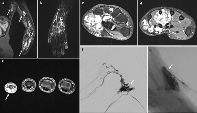
Purpose: To evaluate the safety and outcome of percutaneous sclerotherapy for treating venous malformations (VMs) of the hand.
Materials and methods: A retrospective multicenter trial of 29 patients with VMs primarily affecting the hand, including wrist, carpus, and/or fingers, treated by 81 percutaneous image-guided sclerotherapies using ethanol gel and/or polidocanol was performed. Clinical and imaging findings were assessed to evaluate clinical response, lesion size reduction, and complication rates. Substratification analysis was performed with respect to the Puig's classification, the sclerosing agent, the injected volume of the sclerosant, and to previously performed treatments.
Results: The mean number of procedures per patient was 2.8 (± 2.2). Last follow-up (mean = 9.2 months) revealed a partial relief of symptoms in 78.9% (15/19), while three patients (15.8%) presented symptom-free and one patient (5.3%) with no improvement. Post-treatment imaging revealed an overall objective response rate of 88.9%. Early post-procedural complications occurred after 5/81 sclerotherapies (6.2%) and were entirely resolved by conservative means. Type of VM (Puig's classification) as well as sclerosing agent had no impact on clinical response (p = 0.85, p = 0.11) or complication rates (p = 0.66, p = 0.69). The complication rates were not associated with the sclerosant volume injected (p = 0.76). In addition, no significant differences in clinical success (p = 0.11) or complication rates (p = 0.89) were detected when comparing patients with history of previous treatments compared to therapy-naive patients.
Conclusion: Percutaneous sclerotherapy is both safe and effective for treating VMs of the hand. Even patients with history of previous treatments benefit from further sclerotherapy showing similar low complication rates to therapy-naive patients.
Level of evidence: Level 4, Retrospective study.
Keywords: Hand; Interventional radiology; Sclerotherapy; Upper extremity; Venous malformation.
© 2021. The Author(s).
Conflict of interest statement
The authors declare that they have no conflict of interest.
Figures

References
-
- Lee BB, Baumgartner I, Berlien P, et al. Diagnosis and treatment of venous malformations consensus document of the international union of phlebology (IUP) updated 2013. Int Angiol. 2015;34(2):97–149. - PubMed
Publication types
MeSH terms
Substances
LinkOut - more resources
Full Text Sources
Research Materials
