Review
. 2021 Aug;27(8):2025-2032.
doi: 10.3201/eid2708.210634.

Mycobacterium microti Infections in Free-Ranging Red Deer (Cervus elaphus)

Review

Mycobacterium microti Infections in Free-Ranging Red Deer (Cervus elaphus)

Giovanni Ghielmetti et al. Emerg Infect Dis. 2021 Aug.

Abstract

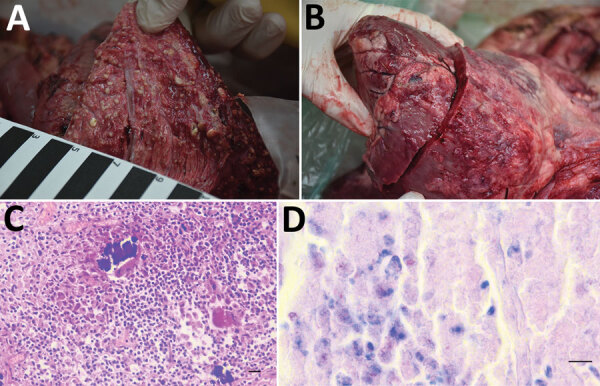
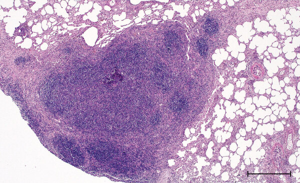
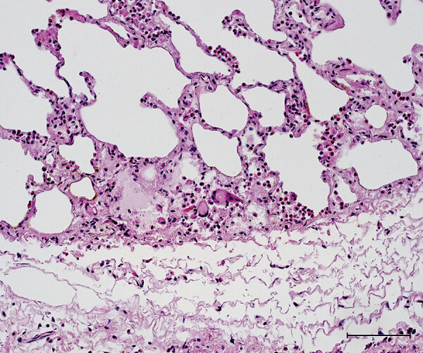
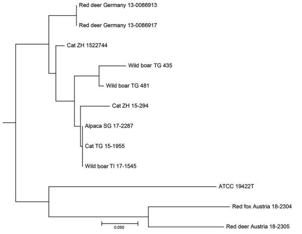
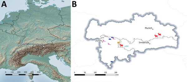
Infections with Mycobacterium microti, a member of the M. tuberculosis complex, have been increasingly reported in humans and in domestic and free-ranging wild animals. At postmortem examination, infected animals may display histopathologic lesions indistinguishable from those caused by M. bovis or M. caprae, potentially leading to misidentification of bovine tuberculosis. We report 3 cases of M. microti infections in free-ranging red deer (Cervus elaphus) from western Austria and southern Germany. One diseased animal displayed severe pyogranulomatous pleuropneumonia and multifocal granulomas on the surface of the pericardium. Two other animals showed alterations of the lungs and associated lymph nodes compatible with parasitic infestation. Results of the phylogenetic analysis including multiple animal strains from the study area showed independent infection events, but no host-adapted genotype. Personnel involved in bovine tuberculosis-monitoring programs should be aware of the fastidious nature of M. microti, its pathogenicity in wildlife, and zoonotic potential.

Keywords: Austria; Cervus elaphus; Germany; MLVA; Mycobacterium microti; Mycobacterium tuberculosis complex; animal hosts; bacteria; disease reservoirs; epidemics; epidemiology; multilocus variable-number tandem-repeat analysis; outbreaks; red deer; respiratory infections; tuberculosis and other mycobacteria; whole-genome sequencing; zoonoses.


Figures

Figure 1
Figure 1
Macroscopic and histopathologic features in the red deer in case 1 in study of tuberculosis caused by Mycobacterium microti in red deer, Austria and Germany. A) Gross picture of the cutting surface of the lungs with severe pyogranulomatous pleuropneumonia with multifocal to confluent cavernous granulomas, 2–10 mm diameter. B) Multifocal to coalescing granulomas 4–25 mm diameter on the surface of the epicardium. C) Chronic multifocal to coalescing pyogranulomatous pneumonia in lungs with central areas of necrosis and mineralization surrounded by numerous epithelioid macrophages and a few multinucleated Langhans giant cells. Single lymphocytes and plasma cells were observed around the periphery and between the granulomas, hematoxylin and eosin stain. Scale bar = 20 μm. D) Numerous macrophages and epithelioid cells containing solitary or multiple acid-fast bacilli. Ziehl Neelsen stain. Scale bar = 10 μm.
Figure 2
Figure 2
Histopathologic features in red deer in case 2 in study of tuberculosis caused by Mycobacterium microti in red deer, Austria and Germany. Lung tissue highly infiltrated by round cells, predominantly lymphocytes and some macrophages, single multinucleated Langhans-type giant cells, hematoxylin and eosin stain. Scale bar = 500 μm.
Figure 3
Figure 3
Histopathologic features in red deer in case 3 in study of tuberculosis caused by Mycobacterium microti in red deer, Austria and Germany. Lung tissue with granulocytic infiltration and some multinucleated Langhans-type giant cells, hematoxylin and eosin stain. Scale bar = 100 μm.
Figure 4
Figure 4
Neighbor-joining tree based on the copy numbers of 24-loci mycobacterial interspersed repetitive unit variable-number tandem-repeat analysis derived from 11 Mycobacterium microti clinical isolates and type strain M. microti Reed ATCC 19422T in study of tuberculosis caused by M. microti in red deer, Austria and Germany. We calculated the tree using the MIRU-VNTRplus server (https://www.miru-vntrplus.org; Appendix) and exported it using MEGAX version 10.11 (https://www.megasoftware.net). Scale bar indicates substitutions per site.
Figure 5
Figure 5
Geographic distribution of tuberculosis cases caused by Mycobacterium microti in different animal species over 8 years from study of tuberculosis caused by M. microti in red deer, Austria and Germany. Central Europe (left) and the region bordering Germany, Austria, and Switzerland (right) are shown. Animals are shaped and colored: red deer, red; cats, pink; wild boar, dark blue; alpaca, orange; and red fox, light blue.

Similar articles

Cited by

References

    1. World Health Organization. Global tuberculosis report 2020. Geneva: World Health Organization. 2020. [cited 2021 Mar 17]. https://www.who.int/publications/i/item/9789240013131
    1. Wells AQ, Robb-Smith AHT. The murine type of tubercle bacillus (the vole acid-fast bacillus); with notes on the morphology of infection by the vole acid-fast bacillus. London: H.M. Stationery Office; 1946.
    1. Kipar A, Burthe SJ, Hetzel U, Rokia MA, Telfer S, Lambin X, et al. Mycobacterium microti tuberculosis in its maintenance host, the field vole (Microtus agrestis): characterization of the disease and possible routes of transmission. Vet Pathol. 2014;51:903–14. 10.1177/0300985813513040 - DOI - PMC - PubMed
    1. Smith NH, Crawshaw T, Parry J, Birtles RJ. Mycobacterium microti: More diverse than previously thought. J Clin Microbiol. 2009;47:2551–9. 10.1128/JCM.00638-09 - DOI - PMC - PubMed
    1. Peterhans S, Landolt P, Friedel U, Oberhänsli F, Dennler M, Willi B, et al. Mycobacterium microti: not just a coincidental pathogen for cats. Front Vet Sci. 2020;7:590037. 10.3389/fvets.2020.590037 - DOI - PMC - PubMed

LinkOut - more resources