. 2021 Sep 9;95(19):e0029921.
doi: 10.1128/JVI.00299-21. Epub 2021 Jul 21.

Targeted Long-Read Sequencing Reveals Comprehensive Architecture, Burden, and Transcriptional Signatures from Hepatitis B Virus-Associated Integrations and Translocations in Hepatocellular Carcinoma Cell Lines

Affiliations

Targeted Long-Read Sequencing Reveals Comprehensive Architecture, Burden, and Transcriptional Signatures from Hepatitis B Virus-Associated Integrations and Translocations in Hepatocellular Carcinoma Cell Lines

Ricardo Ramirez et al. J Virol. .

Abstract

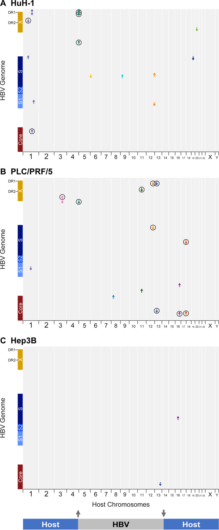
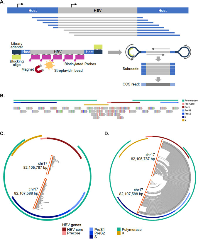
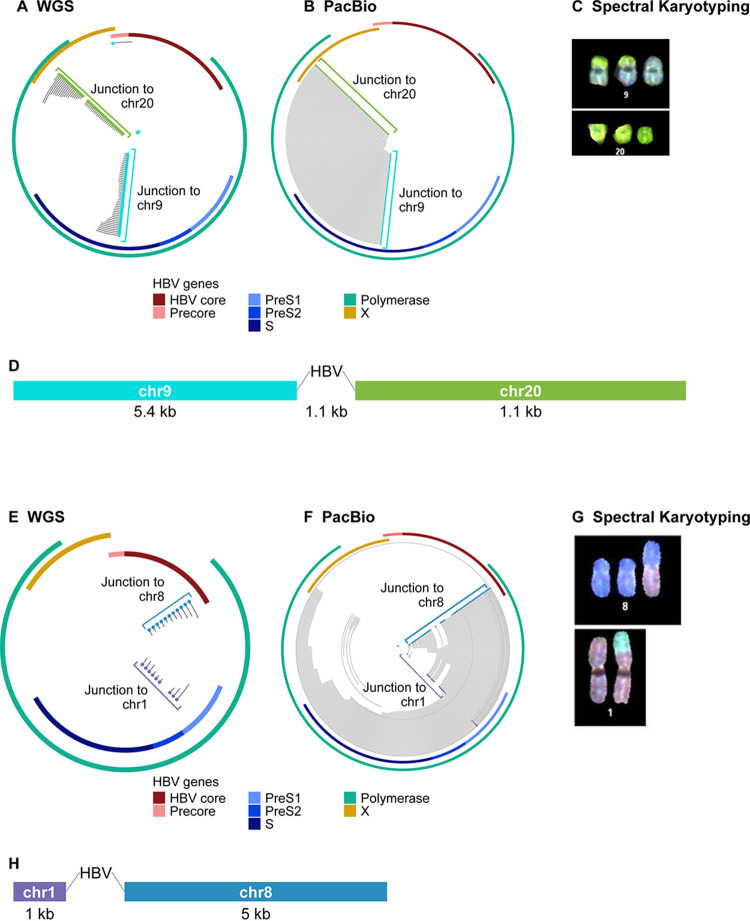
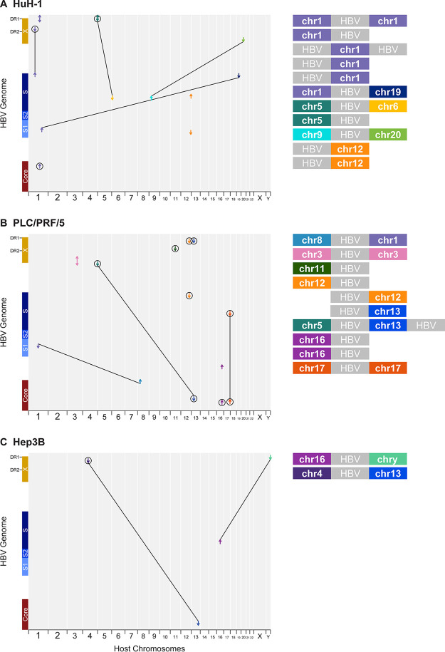
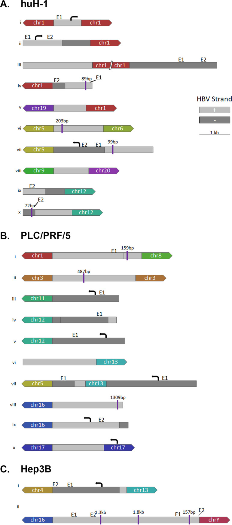
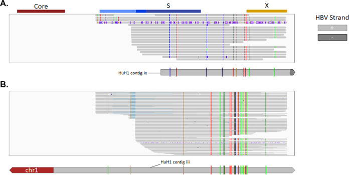
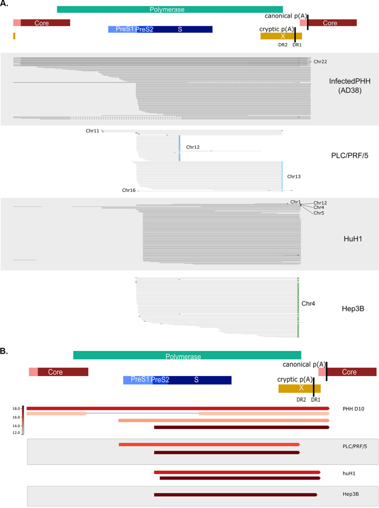
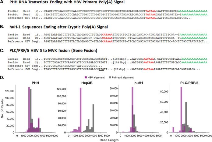
Hepatitis B virus (HBV) can integrate into the chromosomes of infected hepatocytes, creating potentially oncogenic lesions that can lead to hepatocellular carcinoma (HCC). However, our current understanding of integrated HBV DNA architecture, burden, and transcriptional activity is incomplete due to technical limitations. A combination of genomics approaches was used to describe HBV integrations and corresponding transcriptional signatures in three HCC cell lines: huH-1, PLC/PRF/5, and Hep3B. To generate high-coverage, long-read sequencing data, a custom panel of HBV-targeting biotinylated oligonucleotide probes was designed. Targeted long-read DNA sequencing captured entire HBV integration events within individual reads, revealing that integrations may include deletions and inversions of viral sequences. Surprisingly, all three HCC cell lines contain integrations that are associated with host chromosomal translocations. In addition, targeted long-read RNA sequencing allowed for the assignment of transcriptional activity to specific integrations and resolved the contribution of overlapping HBV transcripts. HBV transcripts chimeric with host sequences were resolved in their entirety and often included >1,000 bp of host sequence. This study provides the first comprehensive description of HBV integrations and associated transcriptional activity in three commonly utilized HCC-derived cell lines. The application of novel methods sheds new light on the complexity of these integrations, including HBV bidirectional transcription, nested transcripts, silent integrations, and host genomic rearrangements. The observation of multiple HBV-associated chromosomal translocations gives rise to the hypothesis that HBV is a driver of genetic instability and provides a potential new mechanism for HCC development. IMPORTANCE HCC-derived cell lines have served as practical models to study HBV biology for decades. These cell lines harbor multiple HBV integrations and express only HBV surface antigen (HBsAg). To date, an accurate description of the integration burden, architecture, and transcriptional profile of these cell lines has been limited due to technical constraints. We have developed a targeted long-read sequencing assay that reveals the entire architecture of integrations in these cell lines. In addition, we identified five chromosomal translocations with integrated HBV DNA at the interchromosomal junctions. Incorporation of long-read transcriptome sequencing (RNA-Seq) data indicated that many integrations and translocations were transcriptionally silent. The observation of multiple HBV-associated translocations has strong implications regarding the potential mechanisms for the development of HBV-associated HCC.

Keywords: HBV; HCC; chromosomal translocation; viral integration.


Figures

FIG 1
FIG 1
Short-read sequencing defines locations and transcriptional activity of integrated HBV in HCC cell lines. (A) huH-1, (B) PLC/PRF/5, and (C) Hep3B cell lines were analyzed by short-read RNA-Seq and WGS. HBV integrations were identified by chimeric HBV-host junctions and their locations mapped as arrows according to the aligned sequences to the HBV genome (y axis) and host genome (x axis). Each arrow color corresponds to an individual human chromosome, and its direction correlates to the orientation of the HBV sequence within the associated chimeric read. Circles indicate junctions that were also detected by RNA-Seq.
FIG 2
FIG 2
Targeted PacBio platform established for HBV integrations and reveals complete architecture of integrated HBV DNA. (A) Schematic of target enrichment for PacBio method. (B) Targeting location of the 37 120-bp biotinylated oligonucleotides designed to enrich integrated HBV sequences. (C and D) Comparison between WGS (C) and targeted PacBio (D) data for the same integration on chromosome 17 in PLC/PRF/5 cells. WGS reads identify HBV-host junction ends, while targeted PacBio resolves the entire integration within single reads. Reads are plotted on circle aligning to the HBV genome. Orange circles indicated chimeric junctions with chromosome 17.
FIG 3
FIG 3
Targeted PacBio identifies HBV-associated chromosomal translocations. WGS and targeted PacBio were performed on DNA from huH-1, PLC/PRF/5, and Hep3B cells. (A) WGS data from huH-1 cells found individual HBV-host junctions within chromosomes 9 (blue dots) and 20 (green dots), with no obvious pairing. (B) Targeted PacBio revealed that these junctions originated from an HBV-associated translocation. (C) Chromosome 9 to 20 translocation was confirmed by spectral karyotyping. (D) Alignment of all reads corresponding to this translocation generated a 12-kbp consensus sequence flanking ∼1,100 bp of HBV. (E) WGS data mapped individual HBV-host junctions within Chr1 (purple dots) and Chr8 (blue dots). (F) Targeted PacBio revealed that these junctions originated from an HBV-associated translocation. (G) This translocation event was confirmed by spectral karyotyping. (H) The consensus sequence revealed a segmented HBV with 722 bp adjoined to Chr1 and 2,238 bp adjoined to Chr8.
FIG 4
FIG 4
Multiple HBV integrations are associated with chromosomal translocations. (A to C) Overview of HBV integration and translocation events in huH-1 (A), PLC/PRF/5 (B), and Hep3B (C) cell lines. Each chimeric junction is noted with an arrow mapped against the host genome on the x axis and HBV genome on the y axis. The direction of each arrow indicates the section of HBV included at the respective junction. Lines connecting two chimeric junctions indicate that reads support pairing of those junctions. Diagonal lines connecting two chimeric junctions indicate HBV-associated translocations. Major contigs from each cell line are shown on the right.
FIG 5
FIG 5
HBV architecture and transcriptional profiles of HBV integrations and translocations in huH-1 and PLC/PRF/5 cells. PacBio Reads from huH-1 (A), PLC/PRF/5 (B), and Hep3B (C) cells were used for de novo assembly of each major integration site. The resulting contigs delineated the sequence structure of integrated HBV sequences, including inversions and deletions. Chimeric reads generated by targeted Iso-Seq were used to assign a transcriptional activity, or lack thereof, to each integration event. E1 and E2 denote know enhancer regions (38, 39).
FIG 6
FIG 6
SNVs resolve nonchimeric HBV RNAs from two unique integration events. (A) In addition to chimeric HBV RNAs, we identified a large number of nonchimeric HBV RNAs in huH-1 cells. The majority of these transcripts resolved into two unique SNV profiles. The first SNV aligned contig ix, shown in Fig. 5 (B) The second SNV aligned with contig iii, shown in Fig. 5.
FIG 7
FIG 7
Iso-Seq differentiates transcripts from integrations through unique 3′ termini. (A) Targeted Iso-Seq libraries were generated from huH-1, PLC/PRF/5, and Hep3B cells and AD38-infected PHHs. Gray lines indicate individual long reads. Colored dots indicate reads are chimeric with host chromosomes. (B) Quantification of each transcript was performed by reducing PCR duplicates using UMIs and then quantifying the number of unique reads assigned to each transcript type.
FIG 8
FIG 8
Three novel transcripts types associated with HBV identified by Iso-Seq. Representative HBV RNA sequence reads from PHH (A), PLC/PRF/5 (B), and huH-1 (C) represent the three distinct transcript types identified. The poly(A) signal is in red, while the poly(A) tail is in green. Breakpoints between HBV sequence and host sequence is marked with a caret (^). In panel B, HBV is fused to the MVK gene and is in frame with MVK’s start codon (underlined). (D) Quantification of the host aligning sequence length from chimeric transcripts. A histogram of HBV aligning read length from huH-1, PLC/PRF/5, Hep3B, and AD38 infected PHH cells is plotted in the purple bar graphs. The corresponding total read length, which corresponds to HBV and host aligning sequence, is plotted in gray bar graphs. PLC/PRF/5 and Hep3B cells both contain many chimeric transcripts with >1,000 nucleotides of host sequence appended to the 3′ end of HBV reads.

References

    1. Furuta M, Tanaka H, Shiraishi Y, Unida T, Imamura M, Fujimoto A, Fujita M, Sasaki-Oku A, Maejima K, Nakano K, Kawakami Y, Arihiro K, Aikata H, Ueno M, Hayami S, Ariizumi SI, Yamamoto M, Gotoh K, Ohdan H, Yamaue H, Miyano S, Chayama K, Nakagawa H. 2018. Characterization of HBV integration patterns and timing in liver cancer and HBV-infected livers. Oncotarget 9:25075–25088. 10.18632/oncotarget.25308. - DOI - PMC - PubMed
    1. Mason WS, Gill US, Litwin S, Zhou Y, Peri S, Pop O, Hong ML, Naik S, Quaglia A, Bertoletti A, Kennedy PT. 2016. HBV DNA integration and clonal hepatocyte expansion in chronic hepatitis B patients considered immune tolerant. Gastroenterology 151:986–998. 10.1053/j.gastro.2016.07.012. - DOI - PMC - PubMed
    1. Tu T, Budzinska MA, Shackel NA, Urban S. 2017. HBV DNA integration: molecular mechanisms and clinical implications. Viruses 9:75. 10.3390/v9040075. - DOI - PMC - PubMed
    1. Summers J, Mason WS. 2004. Residual integrated viral DNA after hepadnavirus clearance by nucleoside analog therapy. Proc Natl Acad Sci U S A 101:638–640. 10.1073/pnas.0307422100. - DOI - PMC - PubMed
    1. Ruan P, Dai X, Sun J, He C, Huang C, Zhou R, Chemin I. 2020. Integration of hepatitis B virus DNA into p21-activated kinase 3 (PAK3) gene in HepG2.2.15 cells. Virus Genes 56:168–173. 10.1007/s11262-019-01725-4. - DOI - PubMed

Publication types

Grants and funding

LinkOut - more resources