. 2021 Sep 3;9(1):e0044021.
doi: 10.1128/Spectrum.00440-21. Epub 2021 Jul 21.

Investigating the Roles of Listeria monocytogenes Peroxidases in Growth and Virulence

Affiliations

Investigating the Roles of Listeria monocytogenes Peroxidases in Growth and Virulence

Monica R Cesinger et al. Microbiol Spectr. .

Abstract

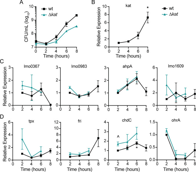
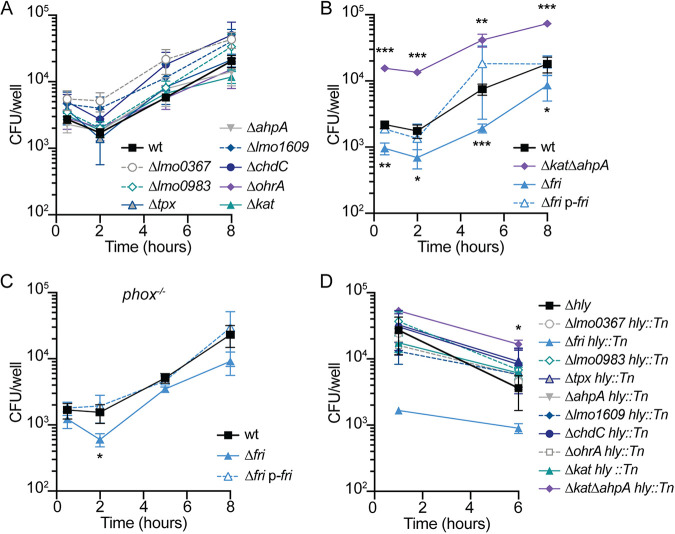
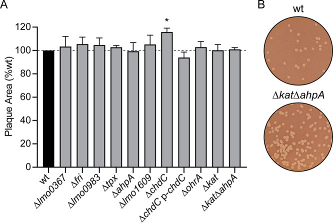
Bacteria have necessarily evolved a protective arsenal of proteins to contend with peroxides and other reactive oxygen species generated in aerobic environments. Listeria monocytogenes encounters an onslaught of peroxide both in the environment and during infection of the mammalian host, where it is the causative agent of the foodborne illness listeriosis. Despite the importance of peroxide for the immune response to bacterial infection, the strategy by which L. monocytogenes protects against peroxide toxicity has yet to be illuminated. Here, we investigated the expression and essentiality of all the peroxidase-encoding genes during L. monocytogenes growth in vitro and during infection of murine cells in tissue culture. We found that chdC and kat were required for aerobic growth in vitro, and fri and ahpA were each required for L. monocytogenes to survive acute peroxide stress. Despite increased expression of fri, ahpA, and kat during infection of macrophages, only fri proved necessary for cytosolic growth. In contrast, the proteins encoded by lmo0367, lmo0983, tpx, lmo1609, and ohrA were dispensable for aerobic growth, acute peroxide detoxification, and infection. Together, our results provide insight into the multifaceted L. monocytogenes peroxide detoxification strategy and demonstrate that L. monocytogenes encodes a functionally diverse set of peroxidase enzymes. IMPORTANCE Listeria monocytogenes is a facultative intracellular pathogen and the causative agent of the foodborne illness listeriosis. L. monocytogenes must contend with reactive oxygen species generated extracellularly during aerobic growth and intracellularly by the host immune system. However, the mechanisms by which L. monocytogenes defends against peroxide toxicity have not yet been defined. Here, we investigated the roles of each of the peroxidase-encoding genes in L. monocytogenes growth, peroxide stress response, and virulence in mammalian cells.

Keywords: heme; oxidative stress; peroxide; redox signaling; virulence.


Figures

FIG 1
FIG 1
Expression of genes encoding putative peroxidases during aerobic growth. (A) Aerobic growth of wt and Δkat strains in shaking flasks was measured by plating for CFU and incubating the plates anaerobically. Data are means and standard errors of the means (SEM) for three biological replicates. (B to D) Relative expression of putative peroxidase-encoding genes over time in both wt and Δkat strains, grown as described for panel A. Expression was normalized to wt expression at 2 h. Data are means and SEM for at least three biological replicates. P values were calculated using a heteroscedastic Student's t test. *, P < 0.05 for expression compared to the wt at 2 h; ^, P < 0.05 for expression in the Δkat mutant compared to the wt at that time point.
FIG 2
FIG 2
Intracellular expression of peroxidase-encoding genes. J774 macrophages were infected with each reporter strain, which expressed rfp from the indicated promoter and constitutive gfp. Cells were infected for 6 h and then analyzed by flow cytometry. The dotted line indicates background RFP fluorescence. Data are means and SEM for three biological replicates. P values were calculated using a heteroscedastic Student's t test comparing each mutant to the wt. **, P < 0.01; ***, P < 0.001.
FIG 3
FIG 3
Aerobic growth of peroxidase mutants. (A) Growth curves of strains that replicate at the same rate as the wt (P > 0.05 at each time point). (B) Growth curves comparing ΔahpA, Δkat, and Δkat ΔahpA mutants. #, P < 0.05 between the Δkat and Δkat ΔahpA strains. (C) Growth curves of strains grown anaerobically (solid lines) or aerobically (dotted lines) overnight before back-diluting into shaking flasks. (D) Strains were grown anaerobically overnight in TSB or BHI and then diluted into TSB with or without exogenous heme (5 μM). In all panels, data are means and SEM for three biological replicates. P values were calculated using a heteroscedastic Student's t test comparing each mutant to the wt grown under the same conditions. *, P < 0.05; **, P < 0.01; ***, P < 0.001.
FIG 4
FIG 4
Acute peroxide toxicity. Bacteria were grown aerobically to mid-log phase before hydrogen peroxide (120 mM) was added. The dotted line indicates the limit of detection (l.o.d.). Data are means and SEM for four biological replicates. P values were calculated using a heteroscedastic Student's t test comparing each mutant to the wt. ***, P < 0.001.
FIG 5
FIG 5
Intracellular survival and growth of peroxidase mutants in IFN-γ-activated BMDMs. (A) Intracellular growth curves of mutant strains that replicated at the same rate as the wt (P > 0.05 at each time point). (B) Intracellular growth curves in activated BMDMs. (C) Intracellular growth curves in activated phox−/− BMDMs, which lack NADPH oxidase. (D) Survival of hly mutants trapped in the vacuoles of activated BMDMs. Although the error bars are too small to be visible, the Δfri hly::Tn strain is not significantly different from the wt at 1 h (P = 0.06). In all panels, data are means and SEM from at least three independent experiments. P values were calculated using a heteroscedastic Student's t test comparing each mutant to the wt grown under the same conditions. *, P < 0.05; **, P < 0.01; ***, P < 0.001.
FIG 6
FIG 6
Intercellular spread of peroxidase mutants. (A) Plaque formation in L2 fibroblasts was evaluated for each strain. Data are means and SEM. The dashed line signifies the 100% level of the wt. (B) Representative images of plaques demonstrating the greater number of plaques formed by the Δkat ΔahpA mutant than the wt.

References

    1. Reniere ML. 2018. Reduce, induce, thrive: bacterial redox sensing during pathogenesis. J Bacteriol 200:903. doi:10.1128/JB.00128-18. - DOI - PMC - PubMed
    1. Nauseef WM, Clark RA. 2019. Intersecting stories of the phagocyte NADPH oxidase and chronic granulomatous disease, p 3–16. In Knaus UG, Leto TL (ed), NADPH oxidases: methods and protocols. Springer, New York, NY. - PubMed
    1. Dinauer MC. 2005. Chronic granulomatous disease and other disorders of phagocyte function. Hematology Am Soc Hematol Educ Program 2005:89–95. doi:10.1182/asheducation-2005.1.89. - DOI - PubMed
    1. van den Berg JM, van Koppen E, Ahlin A, Belohradsky BH, Bernatowska E, Corbeel L, Español T, Fischer A, Kurenko-Deptuch M, Mouy R, Petropoulou T, Roesler J, Seger R, Stasia M-J, Valerius NH, Weening RS, Wolach B, Roos D, Kuijpers TW. 2009. Chronic granulomatous disease: the European experience. PLoS One 4:e5234. doi:10.1371/journal.pone.0005234. - DOI - PMC - PubMed
    1. Imlay JA. 2008. Cellular defenses against superoxide and hydrogen peroxide. Annu Rev Biochem 77:755–776. doi:10.1146/annurev.biochem.77.061606.161055. - DOI - PMC - PubMed

Publication types

LinkOut - more resources