. 2021 Jul 16;54(10):e10891.
doi: 10.1590/1414-431X2020e10891. eCollection 2021.

Juniperus communis extract induces cell cycle arrest and apoptosis of colorectal adenocarcinoma in vitro and in vivo

Affiliations

Juniperus communis extract induces cell cycle arrest and apoptosis of colorectal adenocarcinoma in vitro and in vivo

Wen-Lin Lai et al. Braz J Med Biol Res. .

Abstract

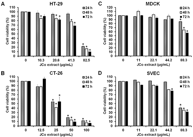
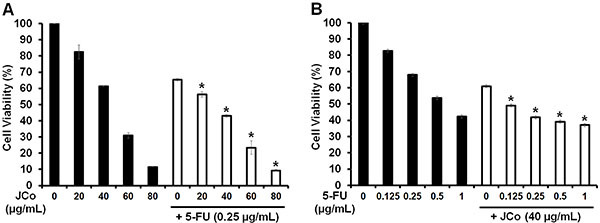
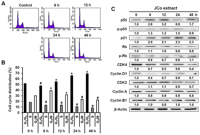
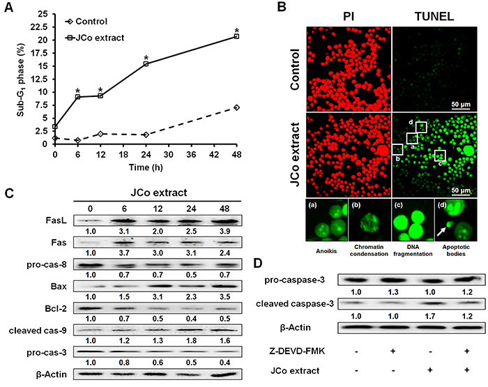
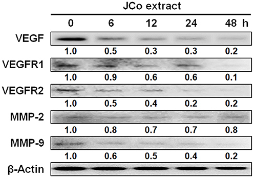
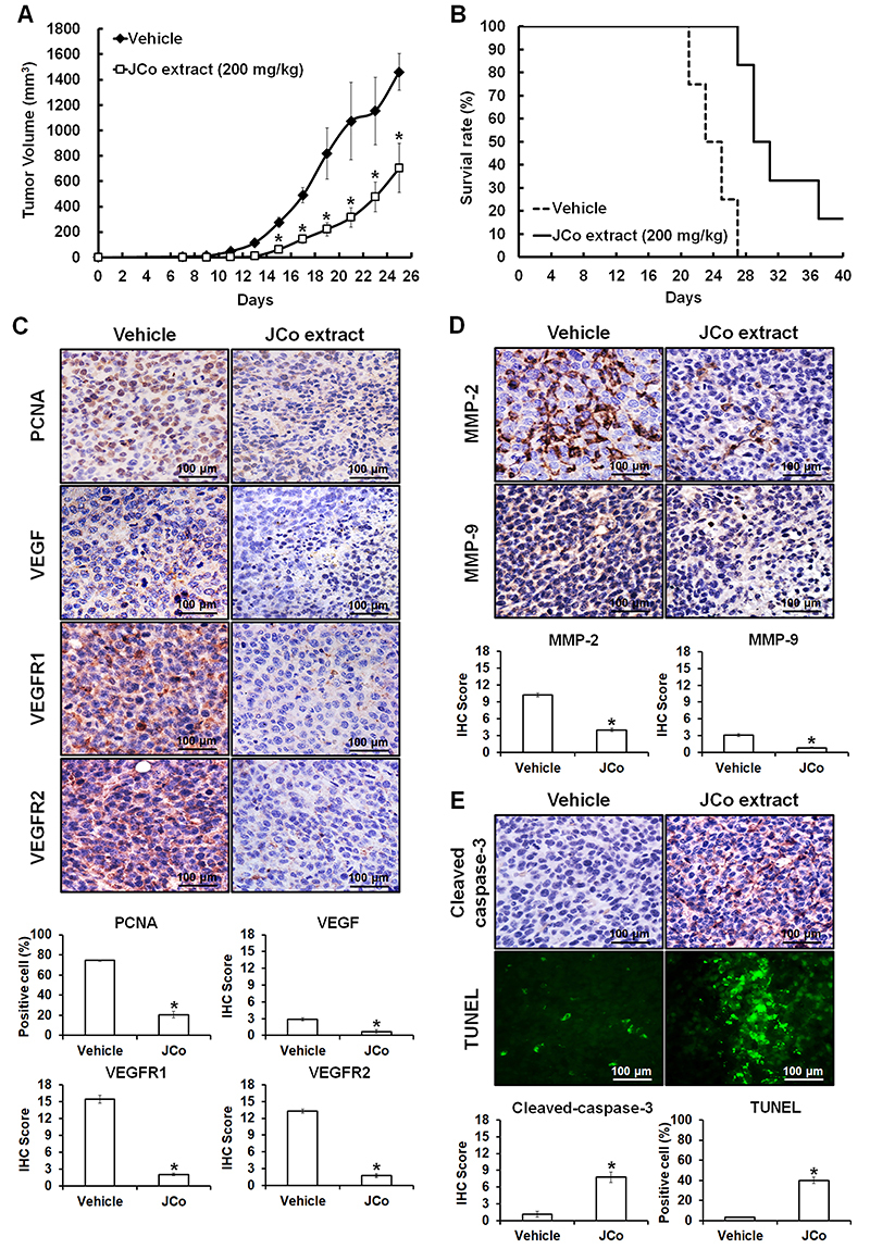
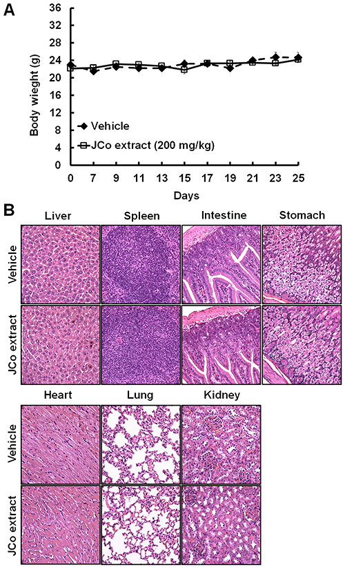
Juniperus communis (JCo) is a well-known traditional Chinese medicinal plant that has been used to treat wounds, fever, swelling, and rheumatism. However, the mechanism underlying the anticancer effect of JCo extract on colorectal cancer (CRC) has not yet been elucidated. This study investigated the anticancer effects of JCo extract in vitro and in vivo as well as the precise molecular mechanisms. Cell viability was evaluated using the MTT assay. Cell cycle distribution was examined by flow cytometry analysis, and cell apoptosis was determined by the terminal deoxynucleotidyl transferase dUTP nick end labeling (TUNEL) assay. Protein expression was analyzed using western blotting. The in vivo activity of the JCo extract was evaluated using a xenograft BALB/c mouse model. The tumors and organs were examined through hematoxylin-eosin (HE) staining and immunohistochemistry. The results showed that JCo extract exhibited higher cytotoxicity against CRC cells than against normal cells and showed synergistic effects when combined with 5-fluorouracil. JCo extract induced cell cycle arrest at the G0/G1 phase via regulation of p53/p21 and CDK4/cyclin D1 and induced cell apoptosis via the extrinsic (FasL/Fas/caspase-8) and intrinsic (Bax/Bcl-2/caspase-9) apoptotic pathways. In vivo studies revealed that JCo extract suppressed tumor growth through the inhibition of proliferation and induction of apoptosis. In addition, there was no obvious change in body weight or histological morphology of normal organs after treatment. JCo extract suppressed CRC progression by inducing cell cycle arrest and apoptosis in vitro and in vivo, suggesting the potential application of JCo extract in the treatment of CRC.


Figures

Figure 1
Figure 1. Effect of Juniperus communis (JCo) extract on growth inhibition in colorectal cancer cell lines. HT-29 (A), CT-26 (B), MDCK (C), and SVEC (D) cells were treated with serial dilutions of JCo extract for 24, 48, and 72 h, and cell viability was evaluated by the 3-(4,5-dimethylthiazol-2-yl)-2,5-diphenyltetrazolium bromide (MTT) assay. Data are reported as means±SD. *P<0.05 vs control (ANOVA).
Figure 2
Figure 2. Juniperus communis (JCo) extract enhances the sensitivity of colorectal cancer cells to 5-fluorouracil (5-FU). HT-29 cells were treated with (A) JCo extract (0, 20, 40, 60, and 80 μg/mL) alone or JCo extract in combination with 0.25 μg/mL 5-FU or with (B) 5-FU (0, 0.125, 0.25, 0.5, and 1 μg/mL) alone or 5-FU in combination with 40 μg/mL JCo extract for 72 h. Cell viability was evaluated by the 3-(4,5-dimethylthiazol-2-yl)-2,5-diphenyltetrazolium bromide (MTT) assay. Data are reported as means±SD. *P<0.05 vs single drug in the combination group. JCo extract in combination with 5-FU displayed a synergistic effect (CI<1) (ANOVA).
Figure 3
Figure 3. Juniperus communis (JCo) extract induces G0/G1 arrest and regulates the expression of cell cycle-associated proteins in HT-29 cells. A, HT-29 cells were treated with 65 μg/mL JCo extract for 0, 6, 12, 24, and 48 h, stained with propidium iodide, and analyzed for FL2 intensity by flow cytometry. B, Cell cycle distribution (G0/G1, S, and G2/M phases) in JCo extract-treated cells was analyzed using Kaluza Flow Cytometry Analysis software. Data are reported as means±SD. *P<0.05 vs control with a significant increase, #P<0.05 vs control with a significant decrease (ANOVA). C, The expression of cell cycle-associated proteins in JCo extract-treated cells was determined by western blotting.
Figure 4
Figure 4. Effects of Juniperus communis (JCo) extract on the extrinsic and intrinsic apoptotic pathways in HT-29 cells. A, The percentage of SubG1 phase cells after JCo extract treatment was analyzed by flow cytometry. Data are reported as means±SD. *P<0.05 vs control (ANOVA). B, Cell apoptosis was determined after treatment with 65 μg/mL JCo extract for 48 h by TUNEL assay (scale bar 50 μm). The apoptotic morphologies included anoikis, chromatin condensation, DNA fragmentation, and the appearance of apoptotic bodies (arrow). C, The protein expression levels of the components of the extrinsic and intrinsic apoptotic pathways in JCo extract-treated cells were analyzed by western blotting. D, HT-29 cells pretreated with 1 μM Z-DEVD-FMK (caspase-3 inhibitor) for 2 h were treated with 65 μg/mL JCo extract for 24 h, and caspase-3 activation was determined by western blotting.
Figure 5
Figure 5. Expression of proteins involved in autocrine angiogenesis and metastasis in Juniperus communis (JCo) extract-treated HT-29 cells. HT-29 cells were treated with 65 μg/mL JCo extract for 0, 6, 12, 24, and 48 h, and the expressions of vascular endothelial growth factor (VEGF), VEGF receptor 1 (VEGFR1), VEGF receptor 2 (VEGFR2), matrix metalloproteinase (MMP)-2, and MMP-9 were analyzed by western blotting.
Figure 6
Figure 6. Effect of Juniperus communis (JCo) extract on the inhibition of CT-26 tumors in a BALB/c mouse model. Subcutaneous tumor-bearing mice were treated with 200 mg/kg JCo extract (sc) once every 2 days for 40 days. A and B, Tumor volumes were calculated once every 2 days, and the mice were sacrificed when the tumor volume exceeded 1500 mm3. C-E, Protein expression levels of proliferating cell nuclear antigen (PCNA), vascular endothelial growth factor (VEGF), VEGF receptor 1 (VEGFR1), VEGF receptor 2 (VEGFR2), matrix metalloproteinase (MMP)-2, MMP-9, and cleaved caspase-3 were detected by immunohistochemistry and scored using the Quickscore method (scale bars 100 μm). JCo extract-induced cell apoptosis was measured using the TUNEL assay. Data are reported as means±SD. *P<0.05 vs vehicle (ANOVA or t-test).
Figure 7
Figure 7. Effects of Juniperus communis (JCo) extract on mouse body weight and vital organs. A, The body weights of tumor-bearing mice were recorded once every 2 days after JCo extract treatment for 40 days. Data are reported as means±SD. B, The vital organs were collected and analyzed by hematoxylin-eosin (HE) staining. No significant differences between the JCo extract and vehicle groups were noted in terms of body weight and histological morphology (scale bars, 100 μm).

Similar articles

Cited by

References

    1. Bray F, Ferlay J, Soerjomataram I, Siegel RL, Torre LA, Jemal A. Global cancer statistics 2018: GLOBOCAN estimates of incidence and mortality worldwide for 36 cancers in 185 countries. CA Can J Clin. 2018;68:394–424. doi: 10.3322/caac.21492. - DOI - PubMed
    1. Grady WM, Carethers JM. Genomic and epigenetic instability in colorectal cancer pathogenesis. Gastroenterology. 2008;135:1079–1099. doi: 10.1053/j.gastro.2008.07.076. - DOI - PMC - PubMed
    1. Yang P, Cartwright C, Chan D, Ding J, Felix E, Pan Y, et al. Anticancer activity of fish oils against human lung cancer is associated with changes in formation of PGE2 and PGE3 and alteration of Akt phosphorylation. Mol Carcinog. 2014;53:566–577. doi: 10.1002/mc.22008. - DOI - PMC - PubMed
    1. Marshall JL, Haller DG, de Gramont A, Hochster HS, Lenz HJ, Ajani JA, et al. Adjuvant therapy for stage II and III colon cancer: consensus report of the international society of gastrointestinal oncology. Gastrointest Cancer Res. 2007;1:146–154. - PMC - PubMed
    1. Smitha KT, Anitha A, Furuike T, Tamura H, Nair SV, Jayakumar R. In vitro evaluation of paclitaxel loaded amorphous chitin nanoparticles for colon cancer drug delivery. Colloids Surf B Biointerfaces. 2013;104:245–253. doi: 10.1016/j.colsurfb.2012.11.031. - DOI - PubMed

Substances