

International Multicenter Analysis of Brain Structure Across Clinical Stages of Parkinson's Disease
- PMID: 34288137
- PMCID: PMC8595579
- DOI: 10.1002/mds.28706
International Multicenter Analysis of Brain Structure Across Clinical Stages of Parkinson's Disease
Abstract
Background: Brain structure abnormalities throughout the course of Parkinson's disease have yet to be fully elucidated.
Objective: Using a multicenter approach and harmonized analysis methods, we aimed to shed light on Parkinson's disease stage-specific profiles of pathology, as suggested by in vivo neuroimaging.
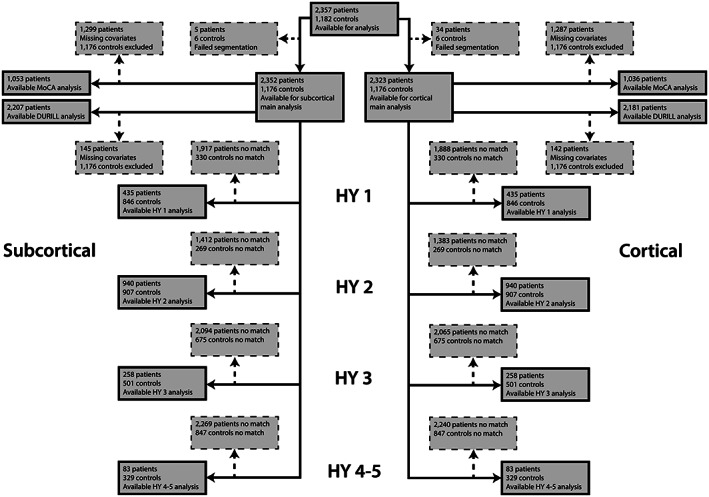
Methods: Individual brain MRI and clinical data from 2357 Parkinson's disease patients and 1182 healthy controls were collected from 19 sources. We analyzed regional cortical thickness, cortical surface area, and subcortical volume using mixed-effects models. Patients grouped according to Hoehn and Yahr stage were compared with age- and sex-matched controls. Within the patient sample, we investigated associations with Montreal Cognitive Assessment score.
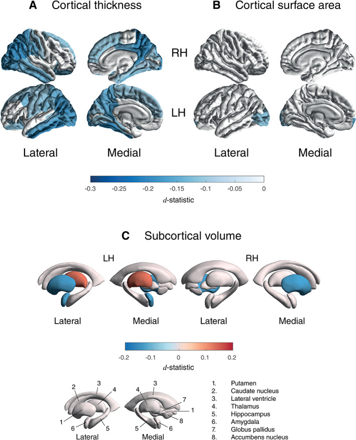
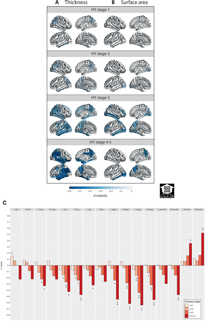
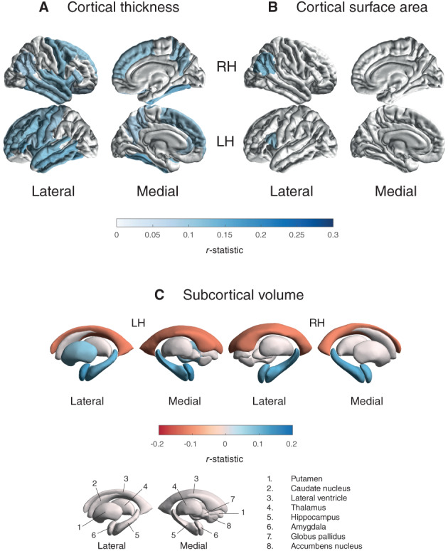
Results: Overall, patients showed a thinner cortex in 38 of 68 regions compared with controls (dmax = -0.20, dmin = -0.09). The bilateral putamen (dleft = -0.14, dright = -0.14) and left amygdala (d = -0.13) were smaller in patients, whereas the left thalamus was larger (d = 0.13). Analysis of staging demonstrated an initial presentation of thinner occipital, parietal, and temporal cortices, extending toward rostrally located cortical regions with increased disease severity. From stage 2 and onward, the bilateral putamen and amygdala were consistently smaller with larger differences denoting each increment. Poorer cognition was associated with widespread cortical thinning and lower volumes of core limbic structures.
Conclusions: Our findings offer robust and novel imaging signatures that are generally incremental across but in certain regions specific to disease stages. Our findings highlight the importance of adequately powered multicenter collaborations. © 2021 The Authors. Movement Disorders published by Wiley Periodicals LLC on behalf of International Parkinson and Movement Disorder Society.
Keywords: ENIGMA; MRI; Parkinson's disease; brain; disease severity.
© 2021 The Authors. Movement Disorders published by Wiley Periodicals LLC on behalf of International Parkinson and Movement Disorder Society.
Figures
Comment in
-
Parkinson's Disease, Premature Mortality, and Amygdala.Mov Disord. 2022 May;37(5):1110-1111. doi: 10.1002/mds.29014. Mov Disord. 2022. PMID: 35587625 No abstract available.
-
Reply to: "Parkinson's Disease, Premature Mortality, and Amygdala".Mov Disord. 2022 May;37(5):1111-1112. doi: 10.1002/mds.29004. Mov Disord. 2022. PMID: 35587628 No abstract available.
References
-
- Poewe W, Seppi K, Tanner CM, et al. Parkinson disease. Nat Rev Dis Primers 2017;3:17013. - PubMed
-
- de Micco R, Russo A, Tessitore A. Structural MRI in idiopathic Parkinson's disease. Int Rev Neurobiol 2018;141:405–438. - PubMed
-
- Hall JM, Lewis SJG. Neural correlates of cognitive impairment in Parkinson's disease: a review of structural MRI findings. Int Rev Neurobiol 2019;144:1–28. - PubMed
