. 2021 Sep 27;60(40):22075-22080.
doi: 10.1002/anie.202103068. Epub 2021 Aug 27.

Cell-Permeable Nanobodies Allow Dual-Color Super-Resolution Microscopy in Untransfected Living Cells

Affiliations

Cell-Permeable Nanobodies Allow Dual-Color Super-Resolution Microscopy in Untransfected Living Cells

Anselm F L Schneider et al. Angew Chem Int Ed Engl. .

Abstract

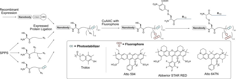
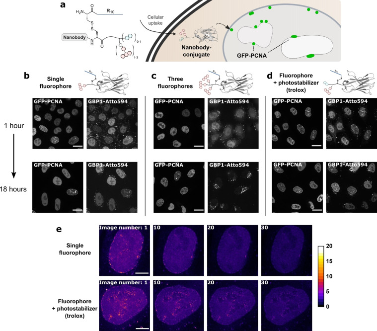
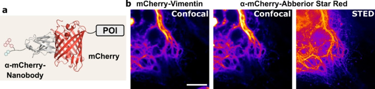
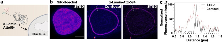
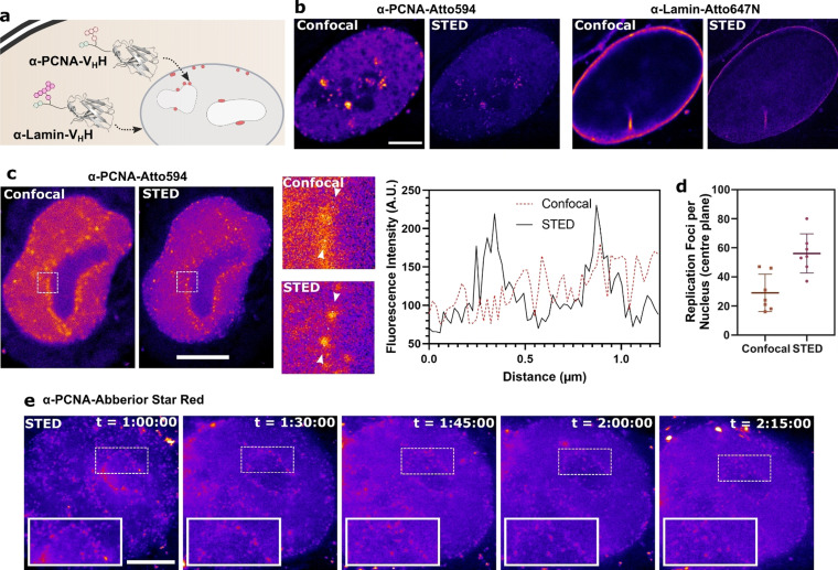
Super-resolution microscopy in living cells can be restricted by the availability of small molecule probes, which only exist against few targets and genetically encoded tags. Here, we expand the applicability of live-cell STED by engineering cell-permeable and highly fluorescent nanobodies as intracellular targeting agents. To ensure bright fluorescent signals at low concentrations we used the concept of intramolecular photostabilization by ligating a fluorophore along with the photostabilizer trolox to the nanobody using expressed protein ligation (EPL). Furthermore, these semi-synthetic nanobodies are equipped with a cleavable cell-penetrating peptide for efficient cellular entry, which enables super-resolution imaging of GFP and mCherry, as well as two endogenous targets, nuclear lamins and the DNA replication and repair protein PCNA. We monitored cell division and DNA replication via confocal and STED microscopy thus demonstrating the utility of these new intracellular tools for functional analysis.

Keywords: STED; cell-penetrating peptides; nanobody; semi-synthesis; super-resolution microscopy.


Conflict of interest statement

The authors declare no conflict of interest.

Figures

Scheme 1
Scheme 1
Semi‐synthetic strategy to obtain fluorescent, cell‐permeable nanobodies. Nanobodies are recombinantly expressed as intein fusion proteins. The intein is cleaved and replaced with a synthetic azide containing peptide, generating attachment sites for a fluorophore and a cell‐penetrating peptide via click chemistry and a disulfide linkage, respectively. The peptides contain either one or three azides and/or the photostabilizer trolox and can be modified with different fluorophores depending on the experimental setup.
Figure 1
Figure 1
Evaluation of nanobody–fluorophore conjugates for super‐resolution microscopy. a) The fluorescent, R10‐functionalized GFP‐binding nanobody GBP1 enters cells after which the R10 peptide is cleaved off. b–d) HeLa kyoto cells expressing GFP–PCNA were treated with 2 μm of the GPB1 nanobody variants. Colocalization with GFP and intracellular stability were assessed by confocal microscopy after 1 and 18 hours post‐treatment of the cells. Scale bars 20 μm. e) STED images from the time series of the nanobody–fluorophore conjugates in living cells. Scale bars 5 μm.
Figure 2
Figure 2
The cell‐permeable mCherry nanobody in STED microscopy. a) Schematic of the Abberior STAR RED‐labeled anti‐mCherry nanobody bound to an mCherry fusion protein. b) STED microscopy of HeLa Kyoto cells transfected with mCherry–Vimentin and treated with 2 μm of the cell‐permeable nanobody. Scale bar 5 μm.
Figure 3
Figure 3
The cell‐permeable lamin nanobody in STED microscopy. a) Schematic of the Atto594‐labeled anti‐lamin nanobody binding to the nuclear lamina. b) STED microscopy of HeLa Kyoto cells treated with 2 μm of the cell‐permeable nanobody with 500 nm SiR‐Hoechst. Scale bar 5 μm. c) Histogram of the normalized fluorescence intensity over a line ROI (see white box in (b)).
Figure 4
Figure 4
The cell‐permeable PCNA nanobody in STED microscopy. a) Schematic of the double labeling experiment using the Atto594‐labeled PCNA nanobody together with the Atto647N‐labeled lamin nanobody binding their nuclear antigens. b) STED microscopy of HeLa Kyoto cells treated with 2 μm of each of the cell‐permeable nanobodies. c) STED and confocal images of a nucleus stained with 2 μm of the cell‐permeable PCNA nanobody. The white squares are shown enlarged. A histogram of the fluorescence intensity was plotted between the white arrows. d) Replication foci were in a single plane of nuclei in confocal and STED microscopy. e) Time‐lapse STED microscopy of a cell in S‐phase stained with the cell‐permeable PCNA nanobody. “t” indicates the timepoints after addition of the nanobody to the cells. Scale bars 5 μm.

References

    1. Sahl S. J., Hell S. W., Jakobs S., Nat. Rev. Mol. Cell Biol. 2017, 18, 685–701. - PubMed
    1. Godin A. G., Lounis B., Cognet L., Biophys. J. 2014, 107, 1777–1784. - PMC - PubMed
    1. None
    1. Westphal V., Rizzoli S. O., Lauterbach M. A., Kamin D., Jahn R., Hell S. W., Science 2008, 320, 246–249; - PubMed
    1. Urban N. T., Willig K. I., Hell S. W., Nagerl U. V., Biophys. J. 2011, 101, 1277–1284; - PMC - PubMed

Publication types

LinkOut - more resources