. 2021 Aug 3;93(30):10424-10434.
doi: 10.1021/acs.analchem.1c02199. Epub 2021 Jul 21.

Exploring the Chemical Space of Protein Glycosylation in Noncovalent Protein Complexes: An Expedition along Different Structural Levels of Human Chorionic Gonadotropin by Employing Mass Spectrometry

Affiliations

Exploring the Chemical Space of Protein Glycosylation in Noncovalent Protein Complexes: An Expedition along Different Structural Levels of Human Chorionic Gonadotropin by Employing Mass Spectrometry

Maximilian Lebede et al. Anal Chem. .

Abstract

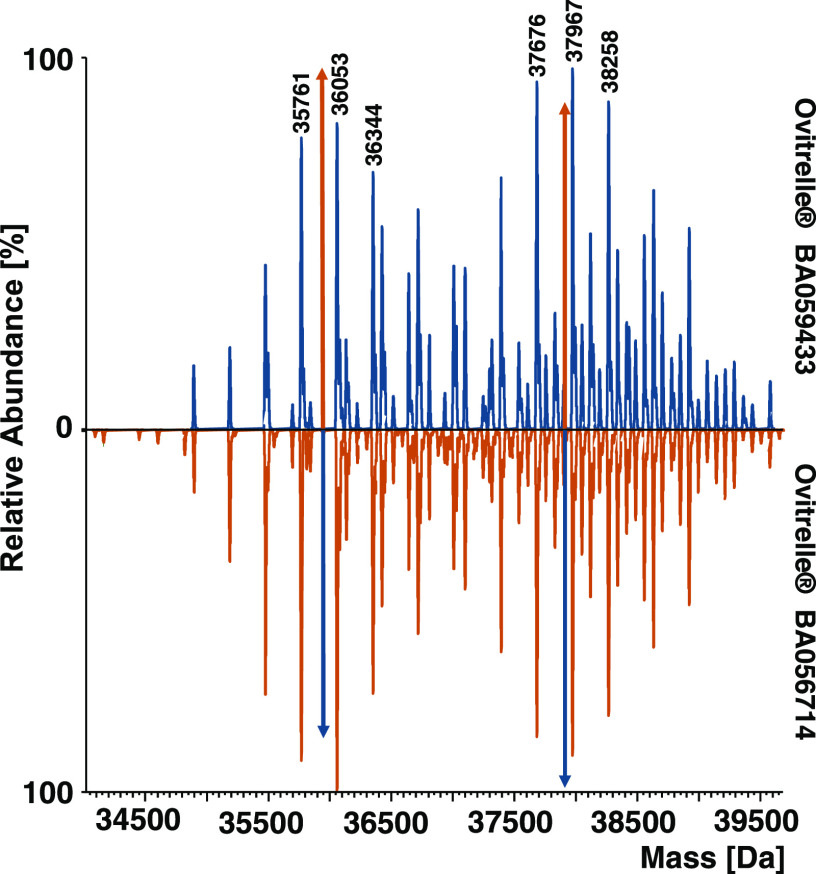
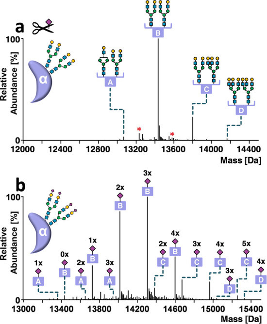
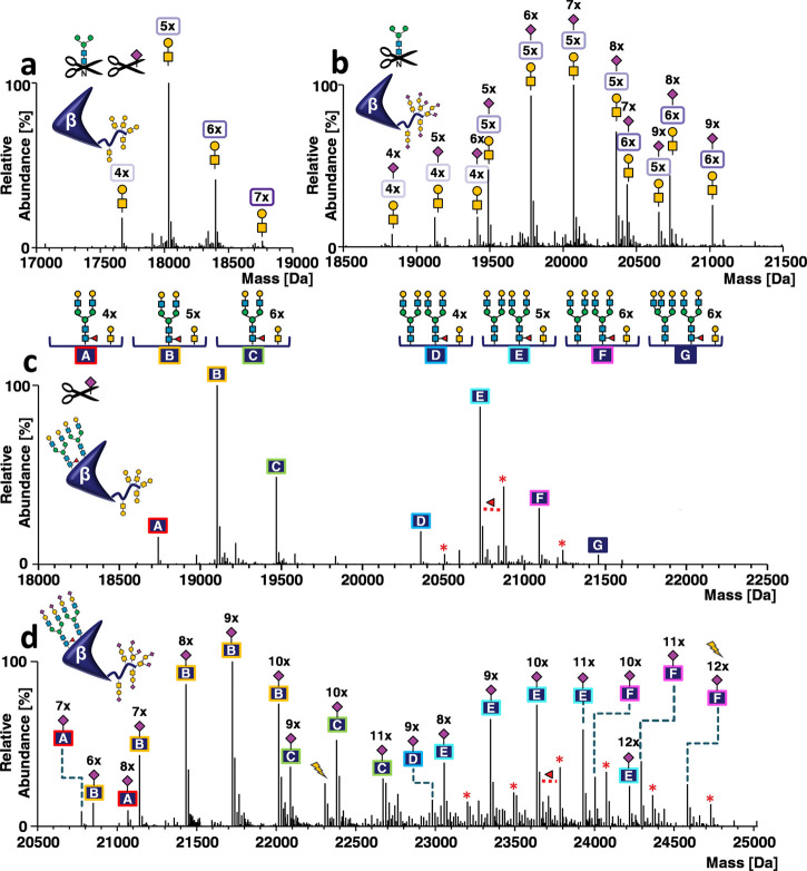
Modern analytical approaches employing high-resolution mass spectrometry (MS) facilitate the generation of a vast amount of structural data of highly complex glycoproteins. Nevertheless, systematic interpretation of this data at different structural levels remains an analytical challenge. The glycoprotein utilized as a model system in this study, human chorionic gonadotropin (hCG), exists as a heterodimer composed of two heavily glycosylated subunits. In order to unravel the multitude of glycoforms of recombinant hCG (drug product Ovitrelle), we combine established techniques, such as released glycan and glycopeptide analysis, with novel approaches employing high-performance liquid chromatography-mass spectrometry (HPLC-MS) to characterize protein subunits and native MS to analyze the noncovalent hCG complex. Starting from the deconvoluted mass spectrum of dimeric hCG comprising about 50 signals, it was possible to explore the chemical space of hCG glycoforms and elucidate the complexity that hides behind just 50 signals. Systematic, stepwise integration of data obtained at the levels of released glycans, glycopeptides, and subunits using a computational annotation tool allowed us to reveal 1031 underlying glycoforms. Additionally, critical quality attributes such as sialylation and core fucosylation were compared for two batches of Ovitrelle to assess the potential product variability.


Conflict of interest statement

The authors declare the following competing financial interest(s): Novartis AG/Sandoz GmbH as well as Thermo Fisher Scientific provide financial support for the Christian Doppler Laboratory for Innovative Tools for Biosimilar Characterization. Rene Hennig is an employee of glyXera GmbH, Magdeburg, Germany. The salaries of Wolfgang Esser-Skala and Therese Wohlschlager are fully funded; Christian G. Huber's salary is partly funded by the Christian Doppler Laboratory for Biosimilar Characterization. The authors declare no other competing financial interest.

Figures

Figure 1
Figure 1
Mirror plot of deconvoluted mass spectra of dimeric hCG of Ovitrelle batches BA059433 and BA056714. Spectra were obtained with an instrument resolution setting of R = 17 500 at m/z 200. Colored arrows represent the peak height of the highest peak in the signal series of the mirrored batch.
Figure 2
Figure 2
Annotation of hCGα glycoforms. Deconvoluted mass spectra of hCGα obtained by HPLC-MS analysis (a) after desialylation or (b) without enzymatic treatment. Glycoforms are indicated by letters (A–D). Red asterisks indicate fucose variants. Numbers above the peaks refer to Neu5Ac residues. Peak lists with all possible glycoform assignments for untreated and desialylated hCGα are available in Supporting Information, Annotations hCGα BA059433. The instrument resolution setting was 140 000 at m/z 200.
Figure 3
Figure 3
Annotation of hCGβ glycoforms. Deconvoluted mass spectra of carbamidomethylated hCGβ obtained by HPLC-MS analysis (a) after sialidase and PNGase F treatment, (b) after PNGase F treatment, (c) after sialidase treatment, and (d) in untreated form. Glycoforms are indicated by letters (A–F). Red asterisks indicate fucose variants. The number of Neu5Ac residues is indicated by the numbers above the peaks. Lightning bolts indicate two peaks that are further discussed in the main text. Peak lists with all possible glycoform assignments for untreated and desialylated hCGβ are available in the Supporting Information. Annotations: hCGβ BA059433. The instrument resolution setting was 140 000 at m/z 200.
Figure 4
Figure 4
Annotation of dimeric hCG glycoforms. (a) Raw mass spectrum of dimeric hCG obtained by native MS. Charge states are indicated. (b) Deconvoluted mass spectrum of dimeric hCG. The glycoforms contributing most to peak abundance are indicated by colored symbols and represent a combination of glycans present on hCGα (A) and hCGβ (B–G). Numbers above these symbols correspond to the number of sialic acids present. The spectrum was obtained with an instrument resolution setting of R = 17 500 at m/z 200.

References

    1. Smith L. M.; Kelleher N. L. Proteoform: A Single Term Describing Protein Complexity. Nat. Methods 2013, 10, 186–187. 10.1038/nmeth.2369. - DOI - PMC - PubMed
    1. Zacchi L. F.; Schulz B. L. N-Glycoprotein Macroheterogeneity: Biological Implications and Proteomic Characterization. Glycoconjugate J. 2016, 33, 359–376. 10.1007/s10719-015-9641-3. - DOI - PubMed
    1. Sandra K.; Vandenheede I.; Sandra P. Modern Chromatographic and Mass Spectrometric Techniques for Protein Biopharmaceutical Characterization. J. Chromatogr. A 2014, 1335, 81–103. 10.1016/j.chroma.2013.11.057. - DOI - PubMed
    1. Beck A.; Wagner-Rousset E.; Ayoub D.; Van Dorsselaer A.; Sanglier S.; Cianférani C. Characterization of Therapeutic Antibodies and Related Products. Anal. Chem. 2012, 85, 715–736. 10.1021/ac3032355. - DOI - PubMed
    1. Wuhrer M.; De Boer A. R.; Deelder A. M. Structural Glycomics Using Hydrophilic Interaction Chromatography (HILIC) with Mass Spectrometry. Mass Spectrom. Rev. 2009, 28, 192–206. 10.1002/mas.20195. - DOI - PubMed

Publication types