

Increased activity of procoagulant factors in patients with small cell lung cancer
- PMID: 34288927
- PMCID: PMC8294523
- DOI: 10.1371/journal.pone.0253613
Increased activity of procoagulant factors in patients with small cell lung cancer
Abstract
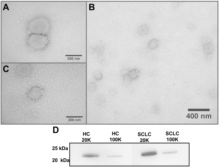
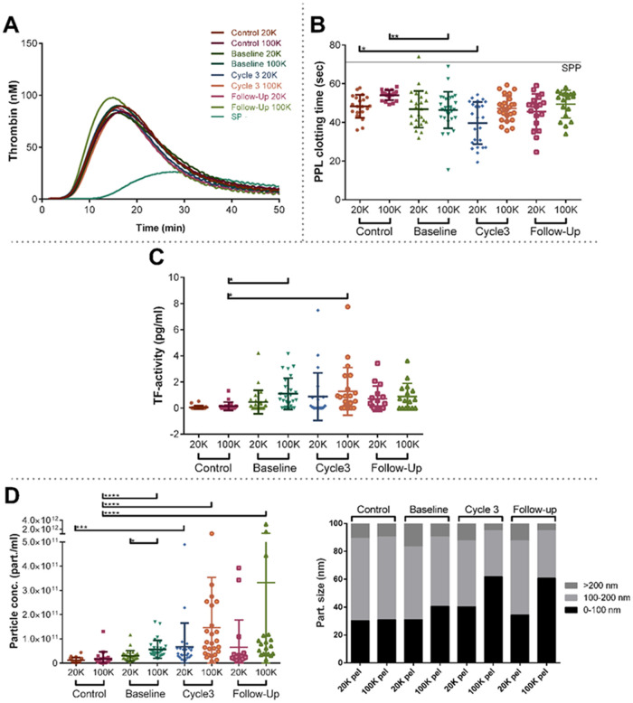
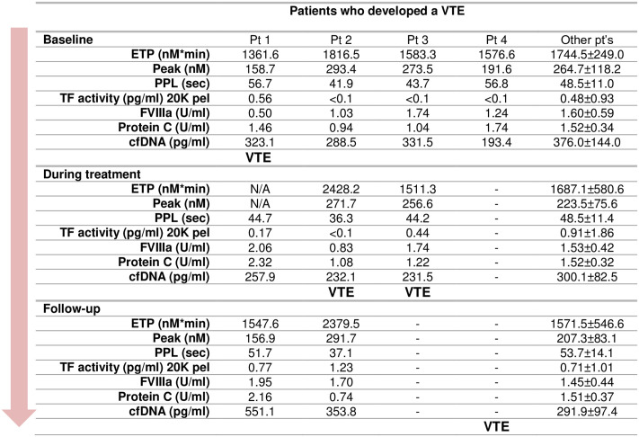
Small cell lung cancer (SCLC) patients have augmented risk of developing venous thromboembolism, but the mechanisms triggering this burden on the coagulation system remain to be understood. Recently, cell-derived microparticles carrying procoagulant phospholipids (PPL) and tissue factor (TF) in their membrane have attracted attention as possible contributors to the thrombogenic processes in cancers. The aims of this study were to assess the coagulation activity of platelet-poor plasma from 38 SCLC patients and to provide a detailed procoagulant profiling of small and large extracellular vesicles (EVs) isolated from these patients at the time of diagnosis, during and after treatment compared to 20 healthy controls. Hypercoagulability testing was performed by thrombin generation (TG), procoagulant phospholipid (PPL), TF activity, Protein C, FVIII activity and cell-free deoxyribonucleic acid (cfDNA), a surrogate measure for neutrophil extracellular traps (NETs). Our results revealed a coagulation activity that is significantly increased in the plasma of SCLC patients when compared to age-related healthy controls, but no substantial changes in coagulation activity during treatment and at follow-up. Although EVs in the patients revealed an increased PPL and TF activity compared with the controls, the TG profiles of EVs added to a standard plasma were similar for patients and controls. Finally, we found no differences in the coagulation profile of patients who developed VTE to those who did not, i.e. the tests could not predict VTE. In conclusion, we found that SCLC patients display an overall increased coagulation activity at time of diagnosis and during the disease, which may contribute to their higher risk of VTE.
Conflict of interest statement
The authors have declared that no competing interests exist.
Figures
Similar articles
-
Extracellular vesicle-associated procoagulant phospholipid and tissue factor activity in multiple myeloma.PLoS One. 2019 Jan 14;14(1):e0210835. doi: 10.1371/journal.pone.0210835. eCollection 2019. PLoS One. 2019. PMID: 30640949 Free PMC article.
-
Coagulation biomarkers and prediction of venous thromboembolism and survival in small cell lung cancer: A sub-study of RASTEN - A randomized trial with low molecular weight heparin.PLoS One. 2018 Nov 9;13(11):e0207387. doi: 10.1371/journal.pone.0207387. eCollection 2018. PLoS One. 2018. PMID: 30412630 Free PMC article. Clinical Trial.
-
Investigation of procoagulant activity in extracellular vesicles isolated by differential ultracentrifugation.J Extracell Vesicles. 2018 Mar 26;7(1):1454777. doi: 10.1080/20013078.2018.1454777. eCollection 2018. J Extracell Vesicles. 2018. PMID: 29696077 Free PMC article.
-
Hypercoagulability and venous thromboembolism in burn patients.Semin Thromb Hemost. 2015 Feb;41(1):43-8. doi: 10.1055/s-0034-1398380. Epub 2015 Jan 15. Semin Thromb Hemost. 2015. PMID: 25590525 Review.
-
Detection of procoagulant imbalance. Modified endogenous thrombin potential with results expressed as ratio of values with-to-without thrombomodulin.Thromb Haemost. 2017 May 3;117(5):830-836. doi: 10.1160/TH16-10-0806. Epub 2017 Feb 23. Thromb Haemost. 2017. PMID: 28229158 Review.
Cited by
-
Circulating microvesicles and exosomes in small cell lung cancer by quantitative proteomics.Clin Proteomics. 2022 Jan 7;19(1):2. doi: 10.1186/s12014-021-09339-5. Clin Proteomics. 2022. PMID: 34996345 Free PMC article.
-
Thromboinflammatory Biomarkers Are Early Predictors of Disease Progression in Non-Small Cell Lung Cancer Patients.Cancers (Basel). 2025 Jun 10;17(12):1932. doi: 10.3390/cancers17121932. Cancers (Basel). 2025. PMID: 40563583 Free PMC article.
-
MPs-ACT, an Assay to Evaluate the Procoagulant Activity of Microparticles.Clin Appl Thromb Hemost. 2023 Jan-Dec;29:10760296231159374. doi: 10.1177/10760296231159374. Clin Appl Thromb Hemost. 2023. PMID: 36843474 Free PMC article.
-
Identifying metabolic alterations in newly diagnosed small cell lung cancer patients.Metabol Open. 2021 Sep 16;12:100127. doi: 10.1016/j.metop.2021.100127. eCollection 2021 Dec. Metabol Open. 2021. PMID: 34585134 Free PMC article.
-
Plasma H3Cit-DNA Discriminates Between Cancer and Inflammation in a Cohort of Patients with Unspecific Cancer Symptoms.Inflammation. 2025 Apr;48(2):760-769. doi: 10.1007/s10753-024-02085-4. Epub 2024 Jun 28. Inflammation. 2025. PMID: 38941006 Free PMC article.
References
Publication types
MeSH terms
Substances
LinkOut - more resources
Full Text Sources
Medical
Miscellaneous
