. 2021 Nov 11;138(19):1830-1842.
doi: 10.1182/blood.2020009192.

Siglec-6 is a novel target for CAR T-cell therapy in acute myeloid leukemia

Affiliations

Siglec-6 is a novel target for CAR T-cell therapy in acute myeloid leukemia

Hardikkumar Jetani et al. Blood. .

Abstract

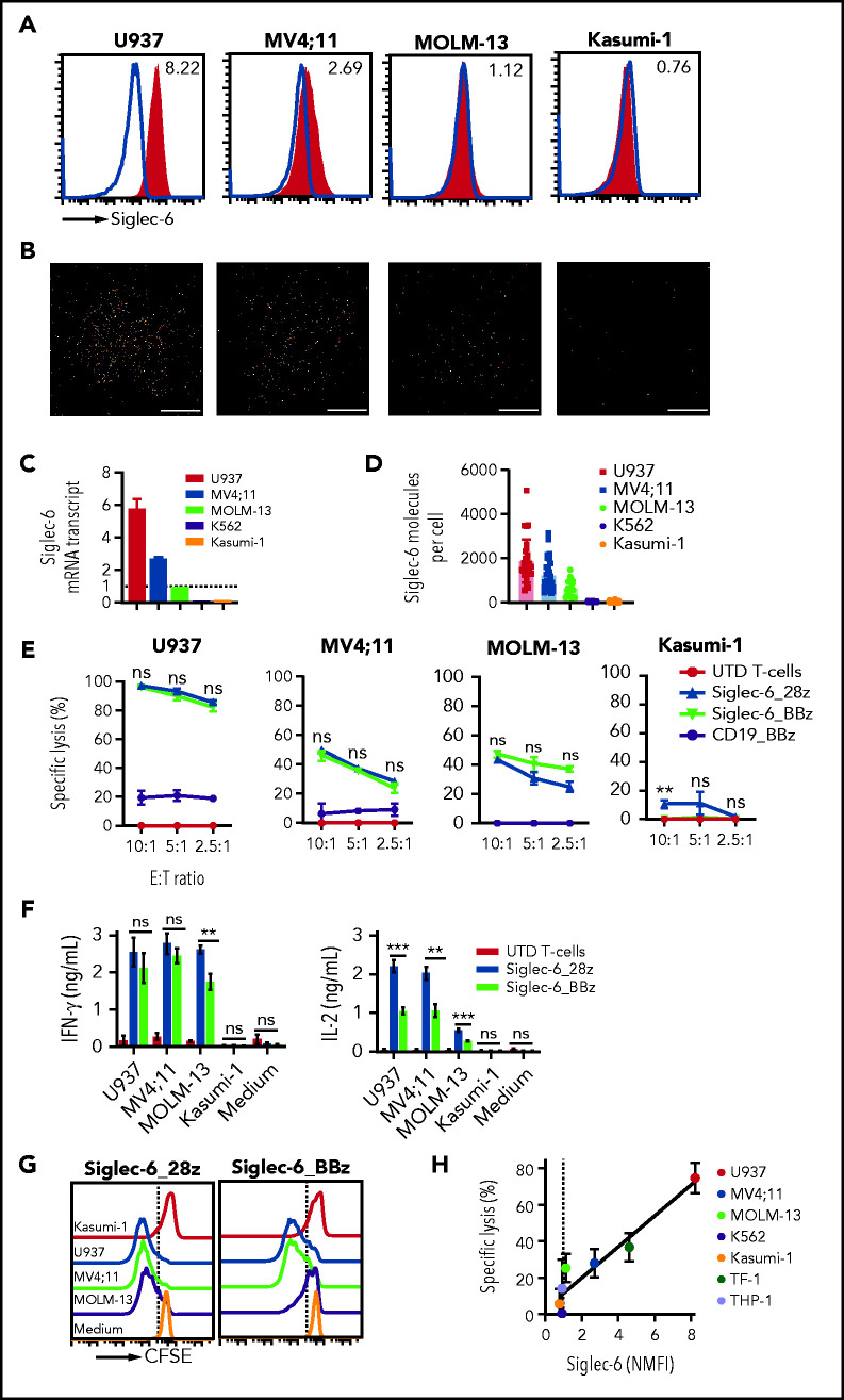
Acute myeloid leukemia (AML) is an attractive entity for the development of chimeric antigen receptor (CAR) T-cell immunotherapy because AML blasts are susceptible to T-cell-mediated elimination. Here, we introduce sialic acid-binding immunoglobulin-like lectin 6 (Siglec-6) as a novel target for CAR T cells in AML. We designed a Siglec-6-specific CAR with a targeting domain derived from the human monoclonal antibody JML-1. We found that Siglec-6 is commonly expressed on AML cell lines and primary AML blasts, including the subpopulation of AML stem cells. Treatment with Siglec-6 CAR T cells confers specific antileukemia reactivity that correlates with Siglec-6 expression in preclinical models, including induction of complete remission in a xenograft AML model in immunodeficient mice (NSG/U937). In addition, we confirmed Siglec-6 expression on transformed B cells in chronic lymphocytic leukemia (CLL), and specific anti-CLL reactivity of Siglec-6 CAR T cells in vitro. Of particular interest, we found that Siglec-6 is not detectable on normal hematopoietic stem and progenitor cells (HSPCs) and that treatment with Siglec-6 CAR T cells does not affect their viability and lineage differentiation in colony-formation assays. These data suggest that Siglec-6 CAR T-cell therapy may be used to effectively treat AML without the need for subsequent allogeneic hematopoietic stem cell transplantation. In mature normal hematopoietic cells, we detected Siglec-6 in a proportion of memory (and naïve) B cells and basophilic granulocytes, suggesting the potential for limited on-target/off-tumor reactivity. The lack of expression of Siglec-6 on normal HSPCs is a key to differentiating it from other Siglec family members (eg, Siglec-3 [CD33]) and other CAR target antigens (eg, CD123) that are under investigation in AML, and it warrants the clinical investigation of Siglec-6 CAR T-cell therapy.


Figures

None
Graphical abstract
Figure 1.
Figure 1.
Siglec-6 is expressed by AML cell lines and Siglec-6 CAR T cells recognize and eliminate AML cells in vitro. (A) Flow cytometric analysis of Siglec-6 expression on AML cell lines (U937, MV4;11, MOLM13, and Kasumi-1). Histograms show staining with anti-Siglec-6 mAb (red) and isotype control antibody (blue). Inset numbers indicate the NMFI calculated by dividing MFI obtained after staining with anti-Siglec-6 mAb by MFI of isotype control. (B) Detection of Siglec-6 expression on AML cell lines using dSTORM super-resolution microscopy. Each representative image depicts Siglec-6 molecules on the basal membrane of a cell. Scale bars represent 5 µm. (C) Real-time qPCR was performed to assess Siglec-6 messenger RNA (mRNA) transcript levels in AML cell lines. Data are normalized to MOLM-13 Siglec-6 mRNA transcript values. (D) Quantification of Siglec-6 expression on AML cell lines by dSTORM super-resolution microscopy. Each point represents a cell. (C-D) Data are representative of 3 independent experiments. (E) Specific cytolytic activity of CD8+ Siglec-6_28z CAR, Siglec-6_BBz CAR, CD19_BBz CAR, and UTD T cells against AML cell lines in a luminescence-based assay (4 hours of coculture). The assay was performed in triplicate wells with 5000 target cells per well. Data are presented as mean ± standard deviation (SD). (F) Enzyme-linked immunosorbent assay (ELISA) was performed to detect interferon-γ (IFN-γ) and interleukin-2 (IL-2) in supernatant obtained after 24-hour coculture of CD8+ Siglec-6_28z CAR, Siglec-6_BBz CAR, or UTD T cells with target cells. T cells and target cells were seeded at a 2:1 E:T ratio in triplicate wells. Data are represented as mean concentration ± SD. (G) Proliferation of CD8+ Siglec-6_28z CAR and Siglec-6_BBz CAR T cells examined by carboxyfluorescein diacetate succinimidyl ester (CFSE) dye dilution after 72 hours of coculture with target cells. The assay was performed in triplicate wells at a (2:1) E:T ratio. Histograms show proliferation of live (7-amino-actinomycin D [7-AAD]) T cells. No exogenous cytokines were added. Data shown in panels E-G are representative of results obtained with CAR and control T-cell lines prepared from at least 5 healthy donors. (H) Correlation between specific lysis by CD8+ Siglec-6_BBz CAR T cells (after 4-hour coculture; 2.5:1 E:T ratio) and Siglec-6 normalized expression on AML cell lines. Simple linear correlation was calculated (R2 = 0.91; P = .0009). test **P < .01; ***P < .001, Student t test. ns, not significant.
Figure 2.
Figure 2.
Siglec-6 is commonly expressed on primary AML blasts and recognized by Siglec-6 CAR T cells. (A) Flow cytometric analysis of Siglec-6 expression on bulk AML blasts and AML stem cells (LSCs) in 3 samples from patients with AML that had high, moderate, and low Siglec-6 expression (Table 1). Histograms show staining with anti–Siglec-6 mAb (red) and isotype control antibody (blue). Inset numbers indicate the NMFI obtained by staining with anti–Siglec-6 mAb and isotype control. (B) Cytolytic activity of CD8+ Siglec-6_BBz CAR and UTD T cells against bulk AML blasts and AML stem cells in a flow cytometry-based 24-hour assay. The experiment was performed in triplicate wells with 10 000 target cells per well. Counting beads were used to quantitate the number of residual live target cells at the end of coculture. (C) Correlation between cytolytic activity by CD8+ Siglec-6_BBz CAR T cells (after 24-hour coculture; 2.5:1 E:T ratio) and Siglec-6 normalized expression on primary AML blasts. Simple linear correlation was calculated (R2 = 0.45; P = .0018).
Figure 3.
Figure 3.
Siglec-6 CAR T cells confer potent antileukemia activity in a xenograft model of AML in vivo. Female NSG mice were inoculated with 2 × 10⁶ U937 AML cells (FLUC+GFP+), and on days 6 and 21, they were treated with 5 × 10⁶ CAR-modified or UTD T cells. T cells were formulated in a 1:1 CD4+:CD8+ ratio. (A) Serial BLI to assess leukemia progression and/or regression. Note the scale indicating upper and lower BLI thresholds at each analysis time point (right). (B) Flow cytometric analysis of peripheral blood on days 10, 14, and 45 to detect T cells and leukemia cells. Human T cells in mouse peripheral blood were defined as 7-AADCD45+CD3+ cells. Leukemia cells were defined as 7-AADCD45+GFP+ cells. (C) Waterfall plot showing change in absolute BLI values between days 6 and 10 after tumor inoculation. (D) BLI values from each treatment group showing tumor progression and regression. BLI values in panels C and D were obtained as photons per second per cm2per sr (p/s/cm2/sr) in regions of interest encompassing the entire body of each mouse. (E) Percentage of leukemic cells detected in bone marrow, spleen, and peripheral blood by flow cytometry at the end of the experiment. NB: Leukemia cells (%) values show data obtained at different time points. Mice from UTD T-cell treatment group were analyzed on day 17, and mice from the Siglec-6 CAR T-cell treatment group were analyzed on day 56. (F) Kaplan-Meier survival analysis for overall survival (left) and progression free survival (right) from different treatment groups. Data shown are representative of results obtained in independent experiments with Siglec-6 CAR T-cell from 2 donors. Mantel-Cox log-rank test ****P < .0001. *P < .05; **P < .01; ****P < .0001, Student t test (B,D-E). Avg, average; d6, day 6.
Figure 4.
Figure 4.
Normal HSPCs are not recognized by Siglec-6 CAR T cells. (A) Flow cytometric analysis of cell surface expression of different CAR target antigens on CD34+CD38 hematopoietic stem cells (HSCs) (left) and CD34+CD38+ hematopoietic progenitor cells (HPCs) (right) from 5 healthy donors. Bar diagrams show NMFI ± standard deviation (SD). Two-way analysis of variance (ANOVA) *P < .05; **P < .01;***P < .001. (B) Quantification of Siglec-6 expression on HSPCs by dSTORM super-resolution microscopy. U937 cells and Siglec-6–negative U937 knockout (KO) cells were used for comparison. Each data point represents a cell. (C) Real-time qPCR was used to assess Siglec-6 mRNA transcripts in CD34+CD38 HSCs and CD34+CD38+ HPCs. Data are normalized to MOLM-13 Siglec-6 mRNA transcripts. AML blasts from patient 24 were included in the analysis as a positive control (supplemental Figure 6). (D) Flow cytometric analysis of Siglec-6 expression on granulocyte colony-stimulating factor–mobilized CD34+CD38 HSCs and CD34+CD38+ HPCs from peripheral blood of 5 healthy donors. Inset numbers indicate the NMFI. (E) Left: percentage of live (7-AAD negative) HSCs after 24-hour coincubation with CD8+ Siglec-6_BBz CAR, CD123_BBz CAR, or UTD T cells. The assay was performed in triplicate wells with 5000 target cells per well. Counting beads were used to quantify the number of residual live HSPCs at the end of co-culture. Data are from 3 independent experiments. Right: colony formation assay was performed with residual live HSPCs after 24 hours of co-incubation with CD8+ Siglec-6_BBz CAR, CD123_BBz CAR, or UTD T cells. Graphs show the absolute number of colonies (mean ± SD) per 55-mm plate as determined by microscopy on day 14 from 3 independent experiments. (F) Flow cytometric analysis of Siglec-6 expression on healthy donor peripheral blood mononuclear cells. Siglec-6 expression by B cells (CD45+CD19+), myeloid cells (CD45+CD33+), T cells (CD45+CD3+CD56), NK cells (CD45+CD56+CD3), and NK T cells (CD45+CD3+CD56+) in 7 healthy donors. Siglec-6 expression by Siglec-6–positive (U937, TF-1, MV4;11, and MOLM-13) and Siglec-6–negative (K562, Kasumi-1) cell lines are plotted for reference. (G) Flow cytometric analysis of Siglec-6 expression on healthy B cells, CD33+ myeloid cells, neutrophils (CD33+CD15+CD16+), and basophils (CD33+CD123+HLA-DR). *P < .05; **P < .01; ***P < .001; ****P < .0001, Student t test. BFU-E, burst-forming unit erythroid; CFU-G, colony-forming unit granulocyte; CFU-M, colony-forming unit macrophage; GEMM, granulocyte, erythrocyte, monocyte, megakaryocyte; GM, granulocyte-macrophage.
Figure 5.
Figure 5.
Siglec-6 CAR T cells recognize malignant B cells in B-CLL. (A) Flow cytometric analysis of Siglec-6 expression on B-CLL cells from 10 patients. Histograms show staining with anti-Siglec-6 mAb (red) and isotype control antibody (blue). Inset numbers indicate the NMFI. (B) Cytolytic activity of CD8+ Siglec-6_BBz CAR, CD19_BBz CAR, and UTD T cells against B-CLL cells in a flow cytometry-based assay. Target cells were seeded in triplicate wells (10 000 cells per well) and were cocultured with effector cells at a 5:1 E:T ratio. Counting beads were used to quantify the number of residual live target cells after 4 hours of coculture (P1, patient 1). (C) Correlation between B-CLL–specific cell lysis by CD8+ Siglec-6_BBz CAR T cells (after 4 hours of coculture, 5:1 E:T ratio) and Siglec-6 expression on primary B-CLL cells. Simple linear correlation was calculated (R2 = 0.54; P = .01). (D) Flow cytometric analysis of Siglec-6 expression on healthy B cells (CD45+CD19+CD5CD20high) from patients with CLL. Left: pooled data on Siglec-6 expression on B-CLL cells from 10 patients and on healthy B-cell subsets from 5 of 10 patients with CLL. The remaining 5 patients did not have enough healthy B cells in the peripheral blood for subset analysis. Right: a representative histogram from patient 3, which shows Siglec-6 expression on healthy immature (CD45+CD19+CD5CD20highCD10+), naïve (CD45+CD19+CD5CD20highCD10CD27), and memory (CD45+CD19+CD5CD20highCD10CD27+) B cells compared with B-CLL cells. (E) Siglec-6 expression on healthy B cells from patients with CLL and healthy donors. *P < .05, Student t test.

Comment in

References

    1. Crocker PR, Varki A. Siglecs in the immune system. Immunology. 2001;103(2):137-145. - PMC - PubMed
    1. Fry TJ, Shah NN, Orentas RJ, et al. . CD22-targeted CAR T cells induce remission in B-ALL that is naive or resistant to CD19-targeted CAR immunotherapy. Nat Med. 2018;24(1):20-28. - PMC - PubMed
    1. Kenderian SS, Ruella M, Shestova O, et al. . CD33-specific chimeric antigen receptor T cells exhibit potent preclinical activity against human acute myeloid leukemia. Leukemia. 2015;29(8):1637-1647. - PMC - PubMed
    1. Kim MY, Yu K-R, Kenderian SS, et al. . Genetic inactivation of CD33 in hematopoietic stem cells to enable CAR T cell immunotherapy for acute myeloid leukemia. Cell. 2018;173(6):1439-1453.e1419. - PMC - PubMed
    1. Haubner S, Perna F, Köhnke T, et al. . Coexpression profile of leukemic stem cell markers for combinatorial targeted therapy in AML. Leukemia. 2019;33(1):64-74. - PMC - PubMed

Publication types