Diagnosis and treatment of nerve injury following venipuncture - A report of two cases
- PMID: 34289298
- PMCID: PMC8342828
- DOI: 10.17085/apm.21010
Diagnosis and treatment of nerve injury following venipuncture - A report of two cases
Abstract
Background: Venipuncture is one of the one of the most commonly performed, minimally-invasive procedures; however, it may lead to peripheral nerve injury. Here, we describe the diagnosis, treatment, and prognosis of two self-reported cases of nerve injury during venipuncture with the aim of drawing attention to possible needle-related nerve injuries.
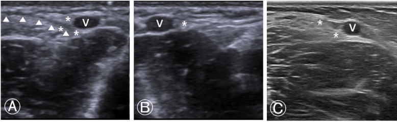
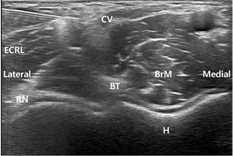
Case: Two anesthesiologists in our hospital experienced an injury of the lateral antebrachial cutaneous branch of the musculocutaneous nerve during venipuncture. Immediately, they underwent ultrasound examinations and nerve blocks with oral medication, resulting in full recovery.
Conclusions: Ultrasonography is important for the early and confirmative diagnosis of a nerve injury during venipuncture, and for immediate treatment with a nerve block. Moreover, it is imperative for both the practitioner and the patient to be aware of the possible complication of nerve injury after venipuncture.
Keywords: Anesthesiologists; Peripheral Nerve Injuries; Ultrasonography; Venipuncture.
Conflict of interest statement
No potential conflict of interest relevant to this article was reported.
Figures
References
-
- Horowitz SH. Peripheral nerve injury and causalgia secondary to routine venipuncture. Neurology. 1994;44:962–4. - PubMed
-
- Newman BH, Waxman DA. Blood donation-related neurologic needle injury: evaluation of 2 years' worth of data from a large blood center. Transfusion. 1996;36:213–5. - PubMed
-
- Kim HJ, Park SK, Park SH. Upper limb nerve injuries caused by intramuscular injection or routine venipuncture. Anesth Pain Med. 2017;12:103–10.
-
- Berry PR, Wallis WE. Venepuncture nerve injuries. Lancet. 1977;309:1236–7. - PubMed
Publication types
LinkOut - more resources
Full Text Sources