. 2022 Feb 1;51(2):20210092.
doi: 10.1259/dmfr.20210092. Epub 2021 Jul 29.

CBCT image artefacts generated by implants located inside the field of view or in the exomass

Affiliations

CBCT image artefacts generated by implants located inside the field of view or in the exomass

Husniye Demirturk Kocasarac et al. Dentomaxillofac Radiol. .

Abstract

Objectives: To compare artefacts in cone-beam computed tomography (CBCT) arising from implants of different materials located either inside the field of view (FOV) or in the exomass, and to test different image-acquisition parameters to reduce them.

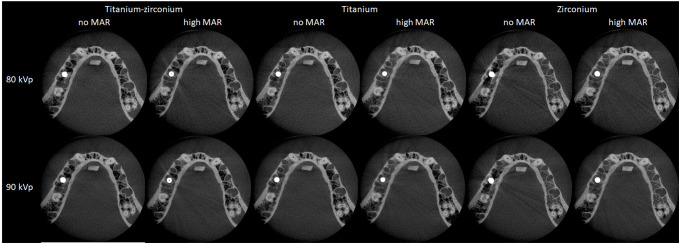
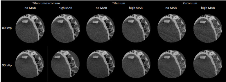
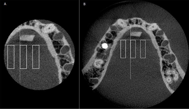
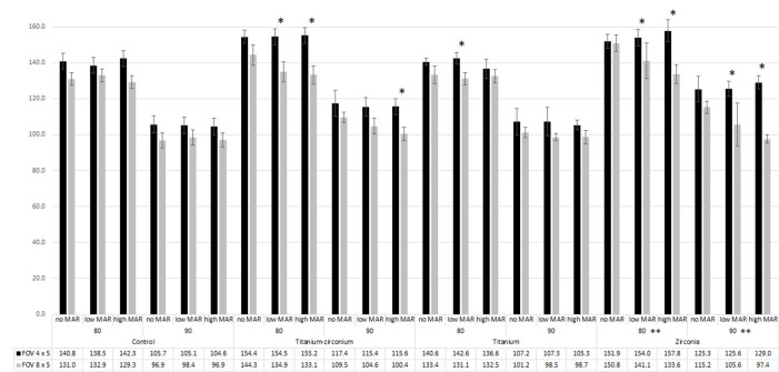
Methods: CBCT scans of a human mandible prepared with either a titanium, titanium-zirconium, or zirconia implant were acquired with the Planmeca ProMax utilizing FOV sizes of 8 × 5 cm and 4 × 5 cm, which placed the implant inside the FOV (8 × 5 cm) or in the exomass (4 × 5 cm). The scanning parameters considered three conditions of metal artefact reduction (MAR), disabled, low, and high, and 2 kVp levels (80 and 90). The standard deviation (SD) of grey values of regions of interest was obtained. The effects of implant material, implant position, MAR condition, kVp level, and their interactions were evaluated by Analysis of Variance (α = 5%).

Results: The zirconia implant produced the highest SD values (more heterogeneous grey values, corresponding to greater artefact expression), followed by titanium-zirconium, and titanium. In general, implants in the exomass produced images with higher SD values than implants inside the FOV. MAR was effective in decreasing SD values, especially from the zirconia implant, only when the implant was inside the FOV. Images with 80 kVp had higher SD values than those with 90 kVp, regardless of the other factors (p < 0.05).

Conclusions: Implants in the exomass lead to greater artefact expression than when they are inside the FOV. Special attention should be paid to scanning parameters that reduce metal-related artefacts, such as MAR activation and increasing kVp. This is especially important with a zirconia implant inside the FOV.

Keywords: Artefacts; CBCT; Titanium; X-Rays; Zirconia.


Conflict of interest statement

Conflict of interest: The authors declare that they have no conflict of interest.

Figures

Figure 1.
Figure 1.
Representative CBCT axial images obtained with a 8 × 5 cm FOV according to the implant material, MAR condition, and kVp level. CBCT, cone-beam CT; FOV, field of view; MAR, metal artefact reduction.
Figure 2.
Figure 2.
Representative CBCT axial images obtained with a 4 × 5 cm FOV according to the implant material, MAR condition, and kVp level. CBCT, cone-beam CT; FOV, field of view; MAR, metal artefact reduction.
Figure 3.
Figure 3.
CBCT axial images indicating the three rectangular regions of interest: A. 4 × 5 cm FOV; B. 8 × 5 cm FOV. White lines are references for the placement of the middle ROI. ROI were established close to the implant, in the middle, and further from the implant, using a line tangent to the alveolar socket of second left premolar and a line perpendicular to the first and tangent to the ERBS block. From the intersection of those lines, the middle ROI was established. The close and further ROIs were drawn 3.6 mm from the middle ROI. . CBCT, cone-beam CT; FOV, field of view; ROI, region of interest.
Figure 4.
Figure 4.
Mean of SD of the grey values according to the implant material, implant position, MAR condition, and kVp level. * indicates significant difference between SD of the grey values when the implant was in the exomass (4 × 5 cm FOV) and inside of the FOV (8 × 5 cm FOV). ** indicates significant difference only for 8 × 5 cm FOV between no MAR and low or high MAR. FOV, field of view; MAR, metal artefact reduction.

References

    1. Schulze R, Heil U, Gross D, Bruellmann DD, Dranischnikow E, Schwanecke U, et al. . Artefacts in CBCT: a review. Dentomaxillofac Radiol 2011; 40: 265–73. doi: 10.1259/dmfr/30642039 - DOI - PMC - PubMed
    1. Freitas DQ, Fontenele RC, Nascimento EHL, Vasconcelos TV, Noujeim M. Influence of acquisition parameters on the magnitude of cone beam computed tomography artifacts. Dentomaxillofac Radiol 2018; 47: 20180151. doi: 10.1259/dmfr.20180151 - DOI - PMC - PubMed
    1. Fontenele RC, Nascimento EH, Vasconcelos TV, Noujeim M, Freitas DQ. Magnitude of cone beam CT image artifacts related to zirconium and titanium implants: impact on image quality. Dentomaxillofac Radiol 2018; 47: 20180021. doi: 10.1259/dmfr.20180021 - DOI - PMC - PubMed
    1. Vasconcelos TV, Bechara BB, McMahan CA, Freitas DQ, Noujeim M. Evaluation of artifacts generated by zirconium implants in cone-beam computed tomography images. Oral Surg Oral Med Oral Pathol Oral Radiol 2017; 123: 265–72. doi: 10.1016/j.oooo.2016.10.021 - DOI - PubMed
    1. Candemil AP, Salmon B, Freitas DQ, Ambrosano GM, Haiter-Neto F, Oliveira ML. Metallic materials in the exomass impair cone beam CT voxel values. Dentomaxillofac Radiol 2018; 47: 20180011. doi: 10.1259/dmfr.20180011 - DOI - PMC - PubMed

Substances