. 2021 Jul 20;36(3):109419.
doi: 10.1016/j.celrep.2021.109419.

Tau oligomer induced HMGB1 release contributes to cellular senescence and neuropathology linked to Alzheimer's disease and frontotemporal dementia

Affiliations

Tau oligomer induced HMGB1 release contributes to cellular senescence and neuropathology linked to Alzheimer's disease and frontotemporal dementia

Sagar Gaikwad et al. Cell Rep. .

Abstract

Aging, pathological tau oligomers (TauO), and chronic inflammation in the brain play a central role in tauopathies, including Alzheimer's disease (AD) and frontotemporal dementia (FTD). However, the underlying mechanism of TauO-induced aging-related neuroinflammation remains unclear. Here, we show that TauO-associated astrocytes display a senescence-like phenotype in the brains of patients with AD and FTD. TauO exposure triggers astrocyte senescence through high mobility group box 1 (HMGB1) release and inflammatory senescence-associated secretory phenotype (SASP), which mediates paracrine senescence in adjacent cells. HMGB1 release inhibition using ethyl pyruvate (EP) and glycyrrhizic acid (GA) prevents TauO-induced senescence through inhibition of p38-mitogen-activated protein kinase (MAPK) and nuclear factor κB (NF-κB)-the essential signaling pathways for SASP development. Despite the developed tauopathy in 12-month-old hTau mice, EP+GA treatment significantly decreases TauO and senescent cell loads in the brain, reduces neuroinflammation, and thus ameliorates cognitive functions. Collectively, TauO-induced HMGB1 release promotes cellular senescence and neuropathology, which could represent an important common pathomechanism in tauopathies including AD and FTD.

Keywords: HMGB1; SASP; aging; astrocytes; cognitive functions; neurodegeneration; neuroinflammation; senescence; tau oligomers; tauopathies.


Conflict of interest statement

Declaration of interests The authors declare no competing interests.

Figures

Figure 1.
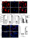
Figure 1.. Astrocytes exhibiting a senescence-like phenotype are associated with TauO in the brain of patients with AD and FTD
(A) Representative immunostaining showing GFAP (green), p16INK4A (red), and TauO (magenta) immunoreactivities and DAPI (blue nuclei) in sections of the frontal cortex from patients with AD and FTD and NDC subjects. In AD and FTD brains, triple immunostaining displays colocalization of p16INK4A and TauO immunoreactivities in GFAP-positive astrocytes. Only a small percentage of cells display p16INK4A and TauO in the NDC brain. Arrows indicate TauO-associated cells exhibiting p16INK4A and GFAP immunoreactivities in AD and FTD. (B and C) Percentage of p16INK4A-positive and GFAP-positive cells, and (C) average numbers of TauO inclusions per 500 μm2 (mean ± SEM; AD n = 8 cases, FTD n = 6 cases, and NDC n = 8 cases; minimum 6–8 images were analyzed from each section). One-way ANOVA followed by Tukey’s post hoc test was used to determine the statistical differences among the groups. Scale bars, 20 μm.
Figure 2.
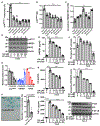
Figure 2.. Nucleo-cytoplasmic translocation and active release of HMGB1 is a hallmark of TauO-induced senescence phenotype in primary astrocytes
(A–F) Upon stimulation with TauO or vehicle for 11 days, astrocytes were examined for HMGB1 translocation and release by immunostaining and flow cytometry analysis, respectively. Arrows point to TauO-induced translocation of HMGB1, a signature of cellular senescence. Scale bars, 50 μm. TauO exposure significantly increased p16INK4A-positive astrocytes (C), the percentage of SA-β-gal-positive astrocytes (D), and cell-cycle arrest (E) p16INK4A staining (F) after 4 days of treatment with or without conditional media (CM) from senescent astrocytes in the presence or absence of α-HMGB1 antibody (4 μg/mL). Representative images showing the relative decrease in number of p16INK4A-positive astrocytes after α-HMGB1 antibody treatment. Scale bar, 50 μm. Data are representative of at least three independent experiments (mean ± SEM). Statistical analyses were measured by unpaired, two-tailed Student’s t test.
Figure 3.
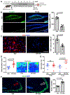
Figure 3.. Inhibition of HMGB1 signaling effectively prevents TauO-induced senescence-like phenotype in cultured astrocytes
Primary astrocytes were cultured in poly-L-lysine (PLL)-coated plates for 48 h and then pretreated with or without HMGB1 release inhibitors EP (10 mM) and/or GA (250 μM) for 30 min followed by treatment with TauO (0.5 μM) for 11 days. (A–C) HMGB1 release inhibition prevents TauO-induced astrocyte senescence, as shown by the decreased percentage of p16INK4A-positive cells and p16INK4A mean fluorescence intensity (MFI) and increased MFI of intracellular HMGB1 by flow cytometry. Representative graphs are shown from a minimum of three to four independent experiments (mean ± SEM). Statistical significance was determined by using one-way ANOVA followed by Tukey’s post hoc test (**p < 0.05; ***p < 0.0001). (D and E) Effect of EP+GA on relative protein levels of p16INK4A, HMGB1, and RAGE was measured by immunoblotting followed by densitometry quantification; β-actin was used as a loading control. The densitometry bar graph is numbered as in blots. Data are shown as mean ± SEM. Statistical significance was determined by using one-way ANOVA followed by Tukey’s post hoc test (**p < 0.05; ***p < 0.0001). (F) Effect of EP+GA on TauO-induced astrocyte senescence was measured by SA-β-gal staining. Pretreatment with EP (10 mM) + GA (250 μM) attenuated TauO-induced astrocytes senescence-like phenotype as shown by decreased SA-β-gal activity. Scale bar, 50 μm. Data are shown as mean ± SEM from four independent experiments in duplicates. Statistical significance was determined using unpaired, two-tailed Student’s t test. (G–I) Conditioned media from the astrocytes culture were used to measure secreted levels of HMGB1, (H) IL-6, and (I) TNF-α using ELISA showing HMGB1 inhibitors effectively inhibit TauO-induced SASP activity. Data are mean ± SEM from at least three independent experiments. Statistical analysis was performed by one-way ANOVA followed by Tukey’s post hoc test. (J) Phosphorylated protein levels of p38 and NF-κB assayed by immunoblotting, followed by densitometry quantification; GAPDH was used as a loading control. Data are shown as mean ± SEM (*p < 0.05; **p < 0.001; ***p < 0.0001).
Figure 4.
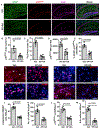
Figure 4.. Treatment with HMGB1 release inhibitors ameliorates tau pathology and cognitive decline in hTau mice
(A) Experimental design for 8-week treatment with HMGB1 release inhibitors EP (80 mg/kg) + GA (20 mg/kg) or vehicle (saline) three times per week, beginning at 12 months of age in hTau mice. (B) Images of thioflavin-S staining and quantification of NFTs (tangles per region of interest) in the hippocampus. Scale bars, 200 μm. Data are shown as the mean ± SEM. (C) Representative immunostaining images showing hyperphosphorylated tau (AT8 immunoreactivities) in the hippocampus of hTau mice treated with either saline or EP+GA (scale bars, 50 μm) and quantification of AT8-positive cells per 500 μm2. Data are the mean ± SEM; unpaired, two-tailed Student’s t test was used to determine the statistical differences. (D) Novel-object recognition task was used to measure the impact of EP+GA treatment on memory in hTau mice. Mice treated with EP+GA show significantly higher discrimination index than vehicle-treated mice; heatmap of representative mice from the vehicle and EP+GA treatment group showing the time spent on exploring old or novel objects. Statistical significance was determined using unpaired, two-tailed Student’s t test. (E) Y-maze spontaneous alternation test: percentage of spontaneous alterations were measured before and after 8 weeks of EP+GA or vehicle treatment. Data are the mean ± SEM; unpaired, two-tailed Student’s t test was used to determine the statistical differences. (F) Immunostaining showing NeuN-positive neuronal cells in the CA3 pyramidal layer of the hippocampus of hTau mice treated with either vehicle or EP+GA. Scale bar, 100 μm. Data are the mean ± SEM; unpaired, two-tailed Student’s t test was used to determine the statistical differences.
Figure 5.
Figure 5.. HMGB1 release inhibitors treatment modulates TauO-associated senescent cells in 12-month-old hTau mice
(A) Representative immunostaining images and quantification of TauO-associated senescent astrocytes in the hippocampus. Representative immunostaining showing GFAP (green), p16INK4A (red), and TauO (magenta) immunoreactivities and DAPI (blue nuclei) in the hippocampus of hTau mice treated with either vehicle or EP+GA. Images display colocalization of p16INK4A and TauO in GFAP-positive astrocytes. (B–E) The graph showing quantitative analysis of p16INK4A area load (B), TauO area load (C), p16INK4A MFI (D), and percentage of p16INK4A-positive and GFAP-positive cells (E) in the hippocampus of hTau mice treated with vehicle (n = 5 mice) and EP+GA (n = 6 mice). Data are shown as mean ± SEM; unpaired, two-tailed Student’s t test was used to determine the statistical differences. Scale bars, 200 μm. (F–M) Immunostaining quantification of cytoplasmic HMGB1-positive cells (F and J), number of γH2AX foci (G and K), p16INK4A-positive cells (H and L), and IL-6-positive cells (I and M) in the cortex of hTau mice treated with vehicle (n = 5 mice) and EP+GA (n = 6 mice). Data are shown as mean ± SEM; unpaired, two-tailed Student’s t test was used to determine the statistical differences. Scale bar, 20 μm.

References

    1. Acosta JC, Banito A, Wuestefeld T, Georgilis A, Janich P, Morton JP, Athineos D, Kang TW, Lasitschka F, Andrulis M, et al. (2013). A complex secretory program orchestrated by the inflammasome controls paracrine senescence. Nat. Cell Biol 15, 978–990. - PMC - PubMed
    1. Andorfer C, Kress Y, Espinoza M, de Silva R, Tucker KL, Barde YA, Duff K, and Davies P (2003). Hyperphosphorylation and aggregation of tau in mice expressing normal human tau isoforms. J. Neurochem 86, 582–590. - PubMed
    1. Andreasen N, Minthon L, Davidsson P, Vanmechelen E, Vanderstichele H, Winblad B, and Blennow K (2001). Evaluation of CSF-tau and CSF-Abeta42 as diagnostic markers for Alzheimer disease in clinical practice. Arch. Neurol 58, 373–379. - PubMed
    1. Arélin K, Kinoshita A, Whelan CM, Irizarry MC, Rebeck GW, Strickland DK, and Hyman BT (2002). LRP and senile plaques in Alzheimer’s disease: colocalization with apolipoprotein E and with activated astrocytes. Brain Res. Mol. Brain Res 104, 38–46. - PubMed
    1. Aucott H, Lundberg J, Salo H, Klevenvall L, Damberg P, Ottosson L, Andersson U, Holmin S, and Erlandsson Harris H (2018). Neuroinflammation in Response to Intracerebral Injections of Different HMGB1 Redox Isoforms. J Innate Immun 10, 215–227. - PMC - PubMed

Publication types

MeSH terms