Review
. 2021 Aug;53(3):350-374.
doi: 10.1080/03602532.2021.1955916. Epub 2021 Aug 25.

Rodent genetic models of Ah receptor signaling

Affiliations
Review

Rodent genetic models of Ah receptor signaling

Rachel H Wilson et al. Drug Metab Rev. 2021 Aug.

Abstract

The aryl hydrocarbon receptor (AHR) is a ligand activated transcription factor that is a member of the PER-ARNT-SIM superfamily of environmental sensors. This receptor has been a molecule of interest for many years in the field of toxicology, as it was originally discovered to mediate the toxic effects of certain environmental pollutants like benzo(a)pyrene and 2,3,7,8-tetrachlorodibenzo-p-dioxin. While all animals express this protein, there is naturally occurring variability in receptor size and responsiveness to ligand. This naturally occurring variation, particularly in mice, has been an essential tool in the discovery and early characterization of the AHR. Genetic models including congenic mice and induced mutations at the Ahr locus have proven invaluable in further understanding the role of the AHR in adaptive metabolism and TCDD-induced toxicity. The creation and examination of Ahr null mice revealed an important physiological role for the AHR in vascular and hepatic development and mediation of the immune system. In this review, we attempt to provide an overview to many of the AHR models that have aided in the understanding of AHR biology thus far. We describe the naturally occurring polymorphisms, congenic models, induced mutations at the Ahr locus and at the binding partner Ah Receptor Nuclear Translocator and chaperone, Ah receptor associated 9 loci in mice, with a brief description of naturally occurring and induced mutations in rats.

Keywords: AHR; ARA9; ARNT; aryl hydrocarbon receptor; gene editing; mouse model; rat model.


Figures

Figure 1.
Figure 1.
AHR signaling pathway. The AHR resides in the cytoplasm as part of a complex bound to two molecules of HSP90, one molecule of P23, and ARA9. Exogenous ligand readily diffuses into the cytoplasm of the cell, where it can bind to the cytosolic AHR complex. The ligand-AHR complex then translocates to the nucleus where the chaperones dissociate. In the nucleus, AHR binds to its obligate partner ARNT to form a heterodimer. The AHR-ARNT dimer recognizes and binds to AHREs located upstream of target genes in the DNA. Binding of AHR-ARNT to AHREs induces transcription of target genes including Cyp1a1, Cyp1a2, Cyp1b1, and Ahrr. The cytochromes P450 (CYP1A1, CYP1A2, and CYP1B1) negatively regulate the pathway by metabolizing the ligands that initiate AHR signaling. The AHRR represses the pathway through interactions with ARNT and binding of the AHRR-ARNT dimer to genomic AHREs.
Figure 2.
Figure 2.
The Ahrb1 (mouse) gene structure and protein map alignment. General domain names are listed in the top row (bHLH, PAS, and TAD), followed by known functions of each region. The bHLH encoding exons and domains are in blue (exons 1 and 2), PAS domains are in green (exons 3–9), and yellow domains represent the TAD (exons 10 and 11). Chromosomal location of the mouse, rat, and human Ahr are listed.
Figure 3.
Figure 3.
Protein maps of mouse AHR naturally occurring alleles and alignments with mouse ARNT. There are four characterized naturally occurring alleles in mice, mAhrb1, mAhrb2, mAhrb3, and mAhrd. The bHLH domain is marked by blue, the PAS domains are shown in green, and the Q rich region of the TAD is shown in yellow. Polymorphisms are shown by the single letter amino acid code on the protein maps. Alignment with the binding partner ARNT is shown to overlap between the bHLH and PAS A domains.
Figure 4.
Figure 4.
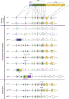
Gene maps for Ahr allelic series. Gene maps of the naturally occurring and induced mutations at the Ahr locus map to a general protein map. Exons encoding for the bHLH are blue, PAS-encoding exons are green, and the TAD is shown in yellow. Exons are numbered and introns are lettered. The light gray region at the 5’ end of exon-1 and the 3’ of exon-11 are the untranslated regions of the exons. Exons that are untranslated as a result of induced mutations and premature stop codons are shown in gray. Alleles are organized by naturally occurring alleles and induced mutations, which is subdivided by the method by which mutations were introduced, either homologous recombination or CRISPR-mediated gene editing. The red arrows represent loxP sites.
Figure 5.
Figure 5.
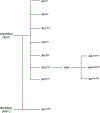
Phylogenetic tree depicting relationships of induced Ahr models. Many of the induced mutations were generated using 129 Sv ESCs. The models generated from these ESCs all harbor the Ahrd allele at the Ahr locus. The Ahrfxneo mouse serves as the parental strain for the Ahrfx, AhrV375A, AhrNG367R, and AhrTer383 models. The AhrCAIR was generated using B6-derived ESCs and harbors the Ahrb1 allele.
Figure 6.
Figure 6.
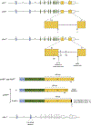
Gene maps of Arnt mutants. The blue exons encode for the bHLH and the green exons encode the PAS domain. The maps are abbreviated at exon-8. The Arntfxneo harbors three loxP (depicted by red triangles) sites, two of which flank exon-6 and one downstream of a NeoPGK cassette. The Arntfx mouse harbors two loxP sites flanking exon-6.
Figure 7.
Figure 7.
Gene maps of Ara9 mutants. The Ara9fxneo harbors two loxP site (depicted by red triangles), two of which flank exons 3 through 6. The Flp sites are shown as yellow triangles and flank a NeoPGK cassette in the intron following exon-6. The Ara9fx mouse harbors two loxP sites flanking exons 3–6.
Figure 8.
Figure 8.
Rat Ahr allele structure and protein maps. A. Gene structure of rat Ahr from Srague-Dawley and Long Evans rats (rAhrSD and rAhrLE) and Han Wistar (rAhrHW). Exons encoding the bHLH are blue, PAS encoding exons are green, and yellow exons encode the TAD. In rAhrSD and rAhrLE rats, the splice donor site (y) splices to the acceptor site (z) at exon 10 and 11 boundaries, respectively. In rAhrHW, there is a G!A mutation in the donor splice site (y), which causes the cell to search elsewhere for cryptic splice sites (a, b, c). One splice site (a) is −129 base pairs from the exon-10 boundary and encodes for the HWdv mRNA. Two other splice sites (b and c) are located in the intron and encode for the HWsiv and HWliv, respectively. B. Protein maps of rat AHR. The rAHRSD encodes a wildtype protein where exon 10 and 11 are joined through the splicing at sites y and z, creating the y-z junction. The rAhrSD animals also encode a Valine (V) at residue 507 in exon 10. The rAhrHW splice variants encode two protein products. The HWdv encodes a protein where cryptic splice site a is used as the new splice donor site and is joined to the z splice donor site at exon-11 (a-z). This results in a protein product that is missing 43 amino acids from exon-10. The HWsiv and HWliv encode for the same protein product which encodes for all of exon-10 and reads through for 7 additional amino acids into the intron (shown in black). Translation is terminated due to the recognition of a new stop codon in the intron which is why HWsic and HWliv encode for the same protein product. Therefore, this protein variant lacks all of exon-11. All rAHRHW proteins harbor an Alanine (A) at residue 507 in exon-10. C. Gene structure of the Ahr null rat. The bHLH was targeted using zinc finger nuclea gene editing and resulted in two alleles, one of which harbors a 2 base pair deletion and one that has a 29 base pair deletion. Both mutations lead to a premature stop codon in exon-2.

References

    1. Abbott BD, Schmid JE, Pitt JA, Buckalew AR, Wood CR, Held GA, & Diliberto JJ (1999). Adverse Reproductive Outcomes in the Transgenic Ah Receptor-Deficient Mouse. Toxicology and Applied Pharmacology, 155(1), 62–70. 10.1006/taap.1998.8601 - DOI - PubMed
    1. Andersson P, McGuire J, Rubio C, Gradin K, Whitelaw ML, Pettersson S, Hanberg A, & Poellinger L (2002). A constitutively active dioxin/aryl hydrocarbon receptor induces stomach tumors. Proceedings of the National Academy of Sciences, 99(15), 9990–9995. 10.1073/pnas.152706299 - DOI - PMC - PubMed
    1. Andreasen EA, Hahn ME, Heideman W, Peterson RE, & Tanguay RL (2002). The Zebrafish (Danio rerio) Aryl Hydrocarbon Receptor Type 1 Is a Novel Vertebrate Receptor. Molecular Pharmacology, 62(2), 234–249. 10.1124/mol.62.2.234 - DOI - PubMed
    1. Anzalone AV, Randolph PB, Davis JR, Sousa AA, Koblan LW, Levy JM, Chen PJ, Wilson C, Newby GA, Raguram A, & Liu DR (2019). Search-and-replace genome editing without double-strand breaks or donor DNA. Nature, 576(7785), 149–157. 10.1038/s41586-019-1711-4 - DOI - PMC - PubMed
    1. Avilla MN, Malecki KMC, Hahn ME, Wilson RH, & Bradfield CA (2020). The Ah Receptor: Adaptive Metabolism, Ligand Diversity, and the Xenokine Model. Chem Res Toxicol, 33(4), 860–879. 10.1021/acs.chemrestox.9b00476 - DOI - PMC - PubMed

Publication types

Substances

LinkOut - more resources