Case Reports
. 2021 Jul 21;14(7):e241285.
doi: 10.1136/bcr-2020-241285.

Spontaneous intracranial hypotension presenting with progressive cognitive decline

Affiliations
Case Reports

Spontaneous intracranial hypotension presenting with progressive cognitive decline

Sanaz Shoja Gharehbagh et al. BMJ Case Rep. .

Abstract

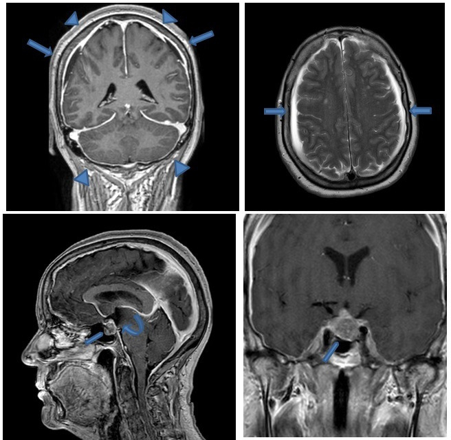
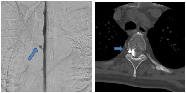
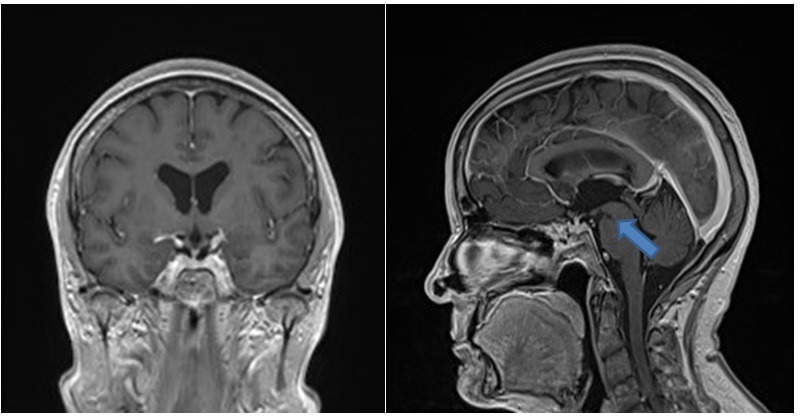
A 63-year-old woman presented with headache, progressive somnolence, neurocognitive decline and urinary incontinence through a year. Medical history was unremarkable except for hypertension and hypercholesterolaemia. Neurological examination was normal. Brain MRI showed findings typical for spontaneous intracranial hypotension (subdural fluid collection, pachymeningeal enhancement, brain sagging) and pituitary tumour. The patient's complaints improved dramatically but temporarily after treatment with each of repeated targeted as well as non-targeted blood patches and a trial with continuous intrathecal saline infusion. Extensive work up including repeated MRI-scans, radioisotope cisternographies, CT and T2-weighted MR myelography could not localise the leakage, but showed minor root-cysts at three levels. Finally, lateral decubitus digital subtraction dynamic myelography with subsequent CT myelography identified a tiny dural venous fistula at the fourth thoracic level. After surgical venous ligation, the patient fully recovered. Awareness of spontaneous dural leaks and their heterogeneous clinical picture are important and demands an extensive workup.

Keywords: headache (including migraines); memory disorders; neuroimaging; pain (neurology).


Conflict of interest statement

Competing interests: None declared.

Figures

Figure 1
Figure 1
Brain MRI with contrast-enhanced T1-weighted sequence shows diffuse smooth pachymeningeal enhancement (arrowheads), bilateral small subdural effusions arrows), and brain sagging with narrowed prepontine cistern, downslope third ventricle (curved arrow) and cerebellar tonsils, small pontomesencephalic angle. Note also pituitary macroadenoma (pentagon).
Figure 2
Figure 2
(A) Right-sided lateral decubitus digital subtraction myelography shows a CSF venous fistula around a perineural cyst on the level Th4/Th5 on the right. (B) Subsequent CT myelography on right lateral decubitus position confirms the CSF venous fistula. CSF, cerebrospinal fluid.
Figure 3
Figure 3
Control brain MRI with contrast enhanced T1-weighted sequence after surgical treatment of the CSF venous fistula shows nearly complete resolution of the signs of SIH. There is only smaller pontomesencephalic angle. CSF, cerebrospinal fluid.

References

    1. Schievink WI. Misdiagnosis of spontaneous intracranial hypotension. Arch Neurol 2003;60:1713. 10.1001/archneur.60.12.1713 - DOI - PubMed
    1. Kranz PG, Malinzak MD, Amrhein TJ, et al. . Update on the diagnosis and treatment of spontaneous intracranial hypotension. Curr Pain Headache Rep 2017;21:37. 10.1007/s11916-017-0639-3 - DOI - PubMed
    1. Schievink WI, Maya MM, Moser F, et al. . Frequency of spontaneous intracranial hypotension in the emergency department. J Headache Pain 2007;8:325–8. 10.1007/s10194-007-0421-8 - DOI - PMC - PubMed
    1. Schievink WI. Spontaneous spinal cerebrospinal fluid leaks and intracranial hypotension. JAMA 2006;295:2286–96. 10.1001/jama.295.19.2286 - DOI - PubMed
    1. Tsai P-H, Fuh J-L, Lirng J-F, et al. . Heavily T2-weighted Mr myelography in patients with spontaneous intracranial hypotension: a case-control study. Cephalalgia 2007;27:929–34. 10.1111/j.1468-2982.2007.01376.x - DOI - PubMed

Publication types