. 2021 Sep;100(3):295-307.
doi: 10.1124/molpharm.121.000280. Epub 2021 Jul 21.

Dynamic Expression of Transient Receptor Potential Vanilloid-3 and Integrated Signaling with Growth Factor Pathways during Lung Epithelial Wound Repair following Wood Smoke Particle and Other Forms of Lung Cell Injury

Affiliations

Dynamic Expression of Transient Receptor Potential Vanilloid-3 and Integrated Signaling with Growth Factor Pathways during Lung Epithelial Wound Repair following Wood Smoke Particle and Other Forms of Lung Cell Injury

Katherine L Burrell et al. Mol Pharmacol. 2021 Sep.

Abstract

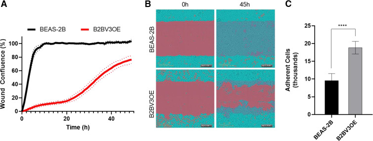
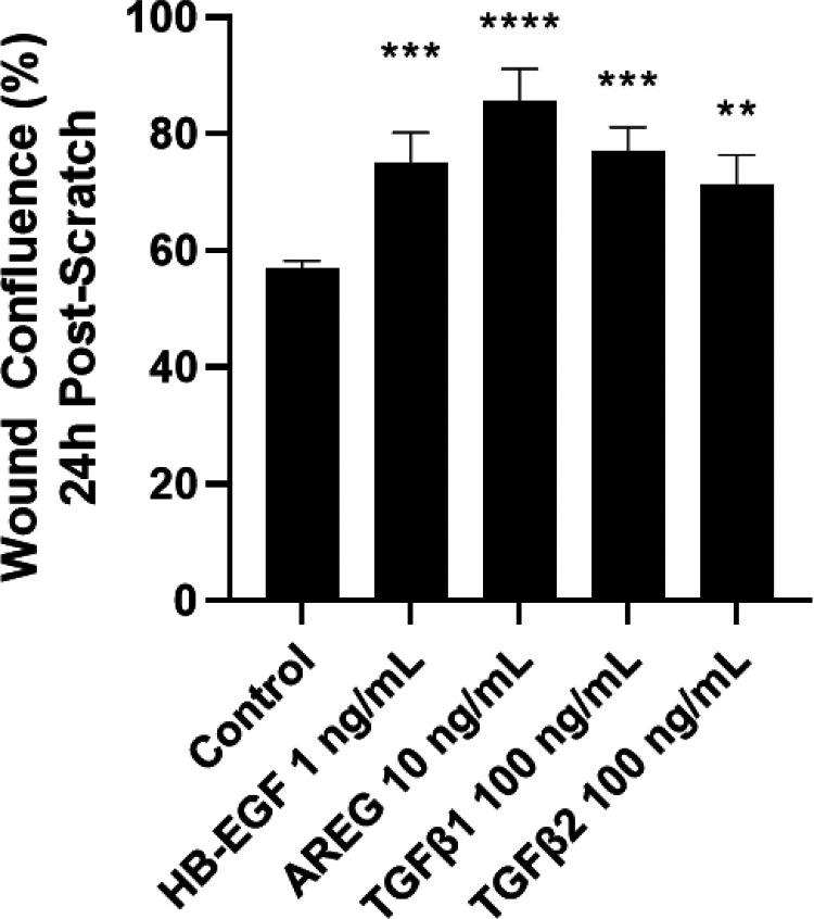
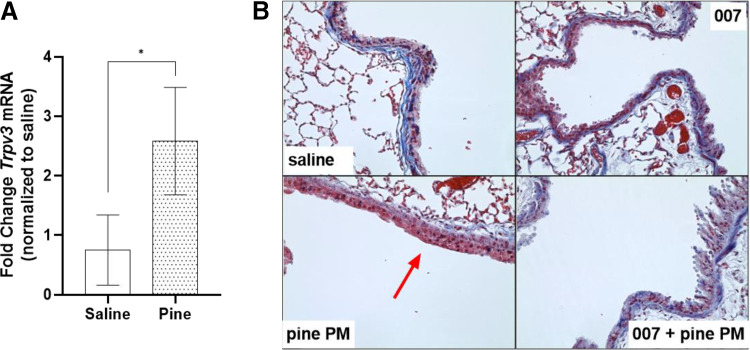
Prior studies revealed increased expression of the transient receptor potential vanilloid-3 (TRPV3) ion channel after wood smoke particulate matter (WSPM) treatment of human bronchial epithelial cells (HBECs). TRPV3 attenuated pathologic endoplasmic reticulum stress and cytotoxicity mediated by transient receptor potential ankyrin-1. Here, the basis for how TRPV3 expression is regulated by cell injury and the effects this has on HBEC physiology and WSPM-induced airway remodeling in mice was investigated. TRPV3 mRNA was rapidly increased in HBECs treated with WSPM and after monolayer damage caused by tryptic disruption, scratch wounding, and cell passaging. TRPV3 mRNA abundance varied with time, and stimulated expression occurred independent of new protein synthesis. Overexpression of TRPV3 in HBECs reduced cell migration and wound repair while enhancing cell adhesion. This phenotype correlated with disrupted mRNA expression of ligands of the epidermal growth factor, tumor growth factor-β, and frizzled receptors. Accordingly, delayed wound repair by TRPV3 overexpressing cells was reversed by growth factor supplementation. In normal HBECs, TRPV3 upregulation was triggered by exogenous growth factor supplementation and was attenuated by inhibitors of growth factor receptor signaling. In mice, subacute oropharyngeal instillation with WSPM also promoted TRPV3 mRNA expression and epithelial remodeling, which was attenuated by TRPV3 antagonist pre- and cotreatment. This latter effect may be the consequence of antagonist-induced TRPV3 expression. These findings provide insights into the roles of TRPV3 in lung epithelial cells under basal and dynamic states, as well as highlight potential roles for TRPV3 ligands in modulating epithelial damage/repair. SIGNIFICANCE STATEMENT: Coordinated epithelial repair is essential for the maintenance of the airways, with deficiencies and exaggerated repair associated with adverse consequences to respiratory health. This study shows that TRPV3, an ion channel, is involved in coordinating repair through integrated repair signaling pathways, wherein TRPV3 expression is upregulated immediately after injury and returns to basal levels as cells complete the repair process. TRPV3 may be a novel target for understanding and/or treating conditions in which airway/lung epithelial repair is not properly orchestrated.


Figures

Fig. 1.
Fig. 1.
TRPV3 expression increased following epithelial injury in vitro. (A) TRPV3 expression in HBEC3-KT cells after four different in vitro models of barrier disruption: 0.076 mg/ml pine PM for 2 hours, disruption of the monolayer by cell passaging, limited trypsin digestion, or mechanical/scratch wounding. (B) Kinetics of TRPV3 mRNA expression after cell passaging injury (left y-axis). The gray fill (right y-axis) represents cell confluence. Data were normalized to 0 hours, noninjured control cells and are presented as the mean ± S.D. from n ≥ 3 replicates. Statistical significance was determined using one-way ANOVA with the Dunnett post-test *P < 0.05, **P < 0.01, ***P < 0.001, ****P < 0.0001. (C) Ca2+ flux assay using the TRPV3 agonist drofenine in HBEC3-KTs cultured for 24 hours postplating (∼20% confluence; white) or 84 hours (100% confluent; gray). Data are presented as mean ± S.D. from n ≥ 3 replicates, and statistical significance was determined using two-way ANOVA with the post hoc Sidak test ****P < 0.0001.
Fig. 2.
Fig. 2.
Effects of transcriptional and translational inhibitors on TRPV3 mRNA expression after epithelial injury in vitro. TRPV3 expression in HBEC3-KT cells was measured 2 hours after passaging injury, with and without treatment with 50 µM ActD (a transcriptional inhibitor) or 100 nM CHX (a protein synthesis inhibitor). Statistical significance was determined using one-way ANOVA with Dunnett’s post-test ***P < 0.001, ****P < 0.0001.
Fig. 3.
Fig. 3.
Stable overexpression of TRPV3 in BEAS-2B HBECs attenuated cell migration and mechanical/scratch wound repair while increasing cell adhesion. (A) BEAS-2B cells (black line) repaired scratch wounds within 6 hours, whereas TRPV3 overexpressing BEAS-2B cells (B2BV3OE, red line) required > 48 hours. Data are shown as the mean ± S.D. (dotted lines) from n = 4 replicates. (B) Live-cell microscopy images (10X) of BEAS-2B and B2BV3OE cells 1 and 40 hours postscratch, with a scratch mask (red) and cell confluence mask (teal) overlaid. Videos of the entire wound repair assay are included as Supplemental Movie 1. (C) Adhesion assay comparing BEAS-2B (black) to B2BV3OE (gray) cells. Data are shown as the mean ± S.D. from n = 8 replicates. Statistical significance was determined using an unpaired t test. ****P < 0.0001.
Fig. 4.
Fig. 4.
Stable overexpression of TRPV3 in BEAS-2B HBECs decreased mRNA expression for multiple EGFR ligands and other canonical growth factor signaling molecules. (A) Log ratio/mean average plot of mRNA sequencing results comparing BEAS-2B and B2BV3OE cells. TRPV3 (green), the growth factors HB-EGF, AREG, NRG1, EPGN, epiregulin, TGFβ1, TGFβ2, and Wnt7a (red), and corresponding growth factor receptors (black) are highlighted. (B) Quantitative analysis of mRNA expression for epiregulin, EPGN, AREG, NRG1, HB-EGF Wnt7a, TGFβ1, and TGFβ2 in confluent B2BV3OE cells. Data were normalized to confluent BEAS-2B cells and shown as the mean ± S.D. from n = 3 replicates. Statistical testing was performed using two-way ANOVA with the post hoc Sidak test. *P > 0.05, **P < 0.01, ***P < 0.001, ****P < 0.0001. (C) Expression of receptors EGFR (ErbB1), ErbB2, ErbB3, FZD5, TGFβRI, and TGFβRII in confluent B2BV3OE cells compared with confluent BEAS-2B cells. Data are shown as the mean ± S.D. from n = 3 replicates performed using two-way ANOVA with the post hoc Sidak test. ****P < 0.0001.
Fig. 5.
Fig. 5.
Addition of downregulated growth factors to TRPV3-overexpressing cells partially rescued the repair deficiency phenotype. Supplementation of B2BV3OE cells with 1 ng/ml HB-EGF, 10 ng/ml AREG, 100 ng/ml TGFβ1, and 100 ng/ml TGFβ2 in growth media increased wound confluence during scratch wound repair compared with vehicle control B2BV3OE cells at 24 hours. Images of scratches after 24 hours are shown in Supplemental Fig. 4. Statistical significance was determined using one-way ANOVA with the Dunnett post-test. **P < 0.01, ***P < 0.001, ****P < 0.0001.
Fig. 6.
Fig. 6.
Changes in EGFR, TGFβ, and Fz receptor ligand expression post–passaging injury, and TRPV3 upregulation by EGFR, TGFβ, and Fz receptor ligand supplementation. (A) Time-dependent upregulation of HB-EGF, AREG, TGFβ1, and Wnt7a mRNA in HBEC3-KT cells compared with 0 hour confluent, noninjured control cells. (B) TRPV3 mRNA expression 2 hours post–passaging injury with and without HB-EGF, AREG, TGFβ1, and Wnt7a supplementation of conditioned media. Data were normalized to 0 hour, noninjured cells and presented as mean ± S.D. from n ≥ 3 replicates. Statistical significance was determined using one-way ANOVA with the Dunnett post-test. *P < 0.05, **P < 0.01, ***P < 0.001, ****P < 0.0001.
Fig. 7.
Fig. 7.
Inhibition of EGFR and growth factor pathway components prevented TRPV3 mRNA upregulation after injury. (A) HBEC3-KT cells were treated with 5 µM solutions of inhibitors of multiple ErbB receptor tyrosine kinase isoforms: ErbB1 (EGFR)-specific inhibitor AG-1478, ErbB pan-inhibitors AZD8931 and Afatinib, and a selective ErbB2 (HER2) inhibitor CP-724 for 2 hours in the cell passaging injury model, followed by analysis of TRPV3 mRNA. (B) Downstream targets of EGFR activation were also inhibited, and TRPV3 expression was subsequently measured 2 hours after cell passaging injury. Inhibitors included 5 µM TWS119 (GSK3β), 20 µM CCT036477 (β-catenin), 10 µM PD169316 (p38 MAPK), and 10 µM SP600125 (JNK). (C) Inhibition of NF-κB (20 µM BMS-345541) and effects on TRPV3 mRNA 2 hours after cell passaging injury. (D) Inhibitors of Wnt7a and Fzd signaling were also used: 10 ng/ml secreted Frizzled-related protein (sFRP) and IWP 2 (Porcupine inhibitor) reduced TRPV3 upregulation after injury. Furthermore, SB 431542 (TGFβRI inhibitor) and 150 µM ITD-1 (TGFβRII inhibitor) were also tested, and SB 431542 treatment prevented TRPV3 expression as well. Values were normalized to vehicle controls and are presented as mean ± S.D. from n ≥ 3 replicates. Statistical significance was determined using one-way ANOVA with the Dunnett post-test. ***P < 0.001, ****P < 0.0001. For Fig. 4C, an unpaired t test was used. ****P < 0.0001.
Fig. 8.
Fig. 8.
Remodeling of, and Trpv3 mRNA upregulation in the airways of mice treated with pine PM. (A) Trpv3 mRNA expression in upper/conducting airway tissue isolated from C57BL/6 mice treated with subacute dosing of 0.5 mg/kg pine PM via OPA. Data represent the mean ± S.D. from n ≥ 3 for each treatment, and statistical significance was determined using an unpaired t test. *P < 0.05. (B) Representative photomicrographs (40X) of trichrome-stained C57BL/6 mouse 1st generation bronchial epithelium after OPA of saline or 0.5 mg/kg pine PM with and without TRPV3 antagonist (007) pre- (1 mg/kg i.p., 1 hour prior) and cotreatment (1 μM OPA). The red arrow highlights remodeled epithelium suggestive of keratinization and epithelial hyperplasia.
Fig. 9.
Fig. 9.
Structurally unique TRPV3 antagonists increased TRPV3 mRNA expression in HBECs and slowed wound repair. (A) Chemical structures of the TRPV3 antagonists 008, 007, and DPTHF. (B) TRPV3 mRNA expression was increased in confluent HBEC3-KT cells after 2 hours treatment with multiple TRPV3 antagonists. Data were normalized to vehicle controls and are presented as the mean ± S.D. from n ≥ 3 replicates. Statistical significance was determined using one-way ANOVA with the Dunnett post-test. **P < 0.01, ****P < 0.0001. Scratch wound repair of HBEC3-KT cells treated with (C) 007 (teal line) or 008 (green line) and (D) DPTHF (red line) compared with untreated controls (black lines). Growth curves are shown as the mean ± S.D. (dotted line) from n = 3 replicates. Live-cell microscopy images (10X) of vehicle versus (E) 300 µM 008 treated cells, and (F) DPTHF treated cells, 0 and 48 hours postscratch. A scratch mask (red) and confluence mask (teal) are overlaid. Videos of the antagonist effect on wound repair are included as Supplemental Movie 2 (008 and 007) and Supplemental Movie 3 (DPTHF).
Fig. 10.
Fig. 10.
Summary schematic of cell signaling pathways that influence, and in turn are influenced by, TRPV3 activity and expression within lung epithelial cells after simulated epithelial injury. Components tested in this study are highlighted as red text. EGFR ligands, TGFβ, and Wnt signaling factors activate signaling cascades including p38 MAPK, NF-κB, GSK3β, and β-catenin to affect TRPV3 expression after injury. It is further suggested that TRPV3 expression and activity in turn affect growth factor expression and shedding to slow repair and to restore proper epithelial homeostasis after injury repair. Other components related to the signaling cascades shown include Ras (a family of small GTPases), Raf (family of kinases), PI3K (Phosphoinositide 3-kinase), MEK (Mitogen-activated protein kinase kinase) and ERK (Extracellular signal-regulated kinase).

References

    1. Abraham CG, Ludwig MP, Andrysik Z, Pandey A, Joshi M, Galbraith MD, Sullivan KD, Espinosa JM (2018) ΔNp63α suppresses TGFB2 expression and RHOA activity to drive cell proliferation in squamous cell carcinomas. Cell Rep 24:3224–3236. - PMC - PubMed
    1. Aijima RWang BTakao TMihara HKashio MOhsaki YZhang J-QMizuno ASuzuki MYamashita Y, et al. (2015) The thermosensitive TRPV3 channel contributes to rapid wound healing in oral epithelia. FASEB J 29:182–192 Federation of American Societies for Experimental Biology. - PubMed
    1. Barrow RE, Wang C-Z, Cox RA, Evans MJ (1992) Cellular sequence of tracheal repair in sheep after smoke inhalation injury. Lung 170:331–338. - PubMed
    1. Borbíró ILisztes ETóth BICzifra GOláh ASzöllosi AGSzentandrássy NNánási PPPéter ZPaus R, et al. (2011) Activation of transient receptor potential vanilloid-3 inhibits human hair growth. J Invest Dermatol 131:1605–1614. - PubMed
    1. Bryant JA, Finn RS, Slamon DJ, Cloughesy TF, Charles AC (2004) EGF activates intracellular and intercellular calcium signaling by distinct pathways in tumor cells. Cancer Biol Ther 3:1243–1249 Taylor & Francis. - PubMed

Publication types

MeSH terms