

Gα15 in early onset of pancreatic ductal adenocarcinoma
- PMID: 34290274
- PMCID: PMC8295279
- DOI: 10.1038/s41598-021-94150-3
Gα15 in early onset of pancreatic ductal adenocarcinoma
Abstract
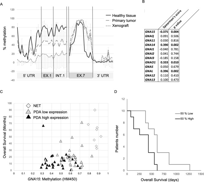
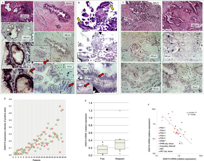
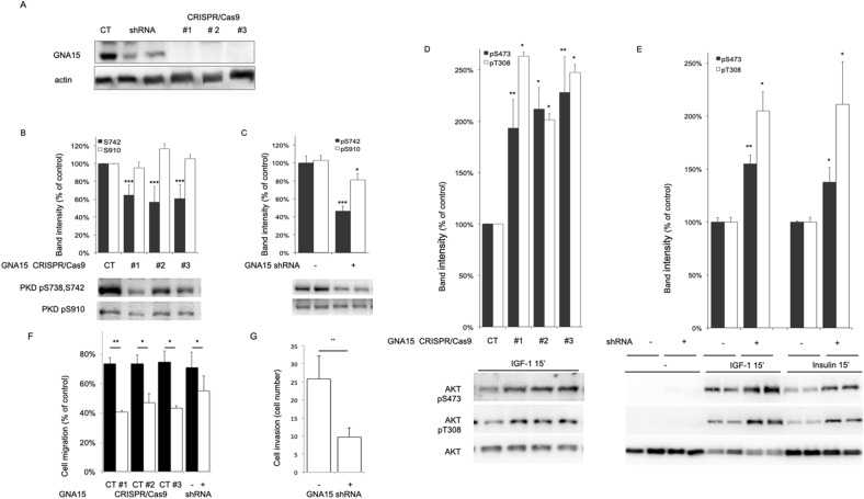
The GNA15 gene is ectopically expressed in human pancreatic ductal adenocarcinoma cancer cells. The encoded Gα15 protein can promiscuously redirect GPCR signaling toward pathways with oncogenic potential. We sought to describe the distribution of GNA15 in adenocarcinoma from human pancreatic specimens and to analyze the mechanism driving abnormal expression and the consequences on signaling and clinical follow-up. We detected GNA15 expression in pre-neoplastic pancreatic lesions and throughout progression. The analysis of biological data sets, primary and xenografted human tumor samples, and clinical follow-up shows that elevated expression is associated with poor prognosis for GNA15, but not any other GNA gene. Demethylation of the 5' GNA15 promoter region was associated with ectopic expression of Gα15 in pancreatic neoplastic cells, but not in adjacent dysplastic or non-transformed tissue. Down-modulation of Gα15 by shRNA or CRISPR/Cas9 affected oncogenic signaling, and reduced adenocarcimoma cell motility and invasiveness. We conclude that de novo expression of wild-type GNA15 characterizes transformed pancreatic cells. The methylation pattern of GNA15 changes in preneoplastic lesions coincident with the release a transcriptional blockade that allows ectopic expression to persist throughout PDAC progression. Elevated GNA15 mRNA correlates with poor prognosis. In addition, ectopic Gα15 signaling provides an unprecedented mechanism in the early steps of pancreas carcinogenesis distinct from classical G protein oncogenic mutations described previously in GNAS and GNAQ/GNA11.
© 2021. The Author(s).
Conflict of interest statement
The authors declare no competing interests.
Figures


Similar articles
-
GNA15 expression in small intestinal neuroendocrine neoplasia: functional and signalling pathway analyses.Cell Signal. 2015 May;27(5):899-907. doi: 10.1016/j.cellsig.2015.02.001. Epub 2015 Feb 17. Cell Signal. 2015. PMID: 25701539
-
KANSL2 and MBNL3 are regulators of pancreatic ductal adenocarcinoma invasion.Sci Rep. 2020 Jan 30;10(1):1485. doi: 10.1038/s41598-020-58448-y. Sci Rep. 2020. PMID: 32001790 Free PMC article.
-
A gene expression signature of epithelial tubulogenesis and a role for ASPM in pancreatic tumor progression.Gastroenterology. 2013 Nov;145(5):1110-20. doi: 10.1053/j.gastro.2013.07.040. Epub 2013 Jul 27. Gastroenterology. 2013. PMID: 23896173
-
Role of miR-30a-3p Regulation of Oncogenic Targets in Pancreatic Ductal Adenocarcinoma Pathogenesis.Int J Mol Sci. 2020 Sep 4;21(18):6459. doi: 10.3390/ijms21186459. Int J Mol Sci. 2020. PMID: 32899691 Free PMC article.
-
MiR-10b inhibits migration and invasion of pancreatic ductal adenocarcinoma via regulating E2F7.J Clin Lab Anal. 2020 Oct;34(10):e23442. doi: 10.1002/jcla.23442. Epub 2020 Jun 26. J Clin Lab Anal. 2020. PMID: 32592206 Free PMC article.
Cited by
-
Histaminergic System and Inflammation-Related Genes in Normal Large Intestine and Adenocarcinoma Tissues: Transcriptional Profiles and Relations.Int J Mol Sci. 2023 Mar 3;24(5):4913. doi: 10.3390/ijms24054913. Int J Mol Sci. 2023. PMID: 36902343 Free PMC article.
-
A novel defined programmed cell death related gene signature for predicting the prognosis of serous ovarian cancer.J Ovarian Res. 2024 Apr 29;17(1):92. doi: 10.1186/s13048-024-01419-y. J Ovarian Res. 2024. PMID: 38685095 Free PMC article.
-
Early-onset gastrointestinal cancer: An epidemiological reality with great significance and implications.World J Gastrointest Oncol. 2024 Mar 15;16(3):583-597. doi: 10.4251/wjgo.v16.i3.583. World J Gastrointest Oncol. 2024. PMID: 38577465 Free PMC article.
-
Tumor-Promoting Role of GNA14 in Colon Cancer Development.Cancers (Basel). 2023 Sep 15;15(18):4572. doi: 10.3390/cancers15184572. Cancers (Basel). 2023. PMID: 37760541 Free PMC article.
-
GNA15 facilitates the malignant development of thyroid carcinoma cells via the BTK-mediated MAPK signaling pathway.Histol Histopathol. 2024 Sep;39(9):1217-1227. doi: 10.14670/HH-18-714. Epub 2024 Jan 19. Histol Histopathol. 2024. PMID: 38333922
References
Publication types
MeSH terms
Substances
Grants and funding
LinkOut - more resources
Full Text Sources
Medical
Research Materials
