. 2021 Jul 21;11(1):14846.
doi: 10.1038/s41598-021-94298-y.

Selective activation of pro-anti-IL-1β antibody enhances specificity for autoinflammatory disorder therapy

Affiliations

Selective activation of pro-anti-IL-1β antibody enhances specificity for autoinflammatory disorder therapy

Wen-Wei Lin et al. Sci Rep. .

Abstract

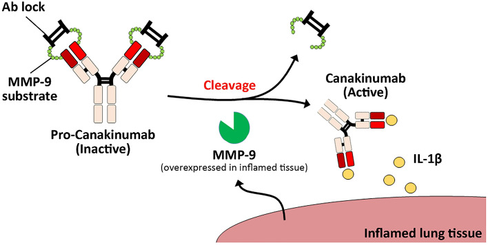
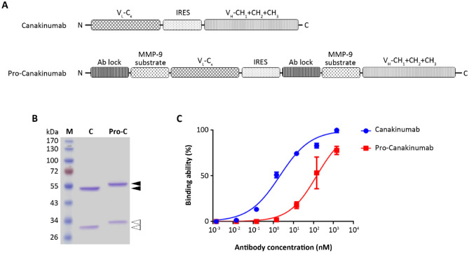
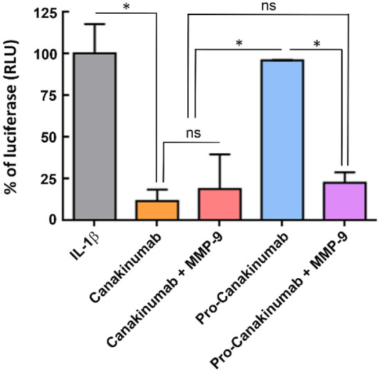
Canakinumab is a fully human monoclonal antibody that specifically neutralizes human interleukin (IL)-1β and has been approved by the US Food and Drug Administration for treating different types of autoinflammatory disorders such as cryopyrin-associated periodic syndrome, tumor necrosis factor receptor-associated periodic syndrome and systemic juvenile idiopathic arthritis. However, long-term systemic neutralization of IL-1β by Canakinumab may cause severe adverse events such as serious upper respiratory tract infections and inflammation, thereby decreasing the quality of life of patients. Here, we used an IgG1 hinge as an Ab lock to cover the IL-1β-binding site of Canakinumab by linking with matrix metalloprotease 9 (MMP-9) substrate to generate pro-Canakinumab that can be specifically activated in the inflamed regions in autoinflammatory diseases to enhance the selectivity and safety of treatment. The Ab lock significantly inhibited the IL-1β-binding by 68-fold compared with Canakinumab, and MMP-9 completely restored the IL-1β neutralizing ability of pro-Canakinumab within 60 min and blocked IL-1β-downstream signaling and IL-1β-regulated genes (i.e., IL-6). It is expected that MMP-9 cleavable and efficient Ab lock will be able to significantly enhance the selective reaction of Canakinumab at the disease site and reduce the on-target toxicities of Canakinumab during systemic circulation, thereby showing potential for development to improve the safety and quality of life of patients with autoinflammatory disorders in the future.


Conflict of interest statement

The authors declare no competing interests.

Figures

Figure 1
Figure 1
Schematic illustration of pro-Canakinumab selective activation by MMP-9 cleavage and specific neutralization of IL-1β at the inflamed region in autoinflammatory diseases. We engineered the human IgG1 hinge as an Ab lock (i.e. EPKSCDKTHTCPPCP) in front of the antigen-binding site of Canakinumab (fully human anti-human IL-1β mAb) by using MMP-9 substrate peptide as linker to generate pro-Canakinumab. After the Ab lock is removed by the MMP-9 expressed in the disease region, the cleaved pro-Canakinumab is expected to be specifically activated, neutralize the local IL-1β antigen and reduce the systemic on-target toxicity during autoinflammatory disease treatment.
Figure 2
Figure 2
Generation and characterization of pro-Canakinumab. (A) Schematic of the Canakinumab (fully human anti-human IL-1β mAb) including (from the N to C terminus) the light chain (VL-Cκ) and heavy chain (VH-CH1 + CH2 + CH3) of Canakinumab that link with an internal ribosomal entry site (IRES). The pro-Canakinumab includes (from the N to C terminus) the IgG1 hinge (Ab lock) in front of the light chain (VL-Cκ) and heavy chain (VH-CH1 + CH2 + CH3) of Canakinumab, respectively, and these two fragments are also joined by an IRES. (B) SDS-PAGE analysis of protein A-purified Canakinumab and pro-Canakinumab: lane 1, molecular weight marker; lane 2, Canakinumab; lane 3, pro-Canakinumab. The black arrow indicates the heavy chain fragments of Canakinumab or pro-Canakinumab, respectively. The white arrow indicates the light chain fragments of Canakinumab or pro-Canakinumab, respectively. (C) The binding ability of Canakinumab (blue circles) and pro-Canakinumab (red squares) was assessed by IL-1β-based ELISA. The percentage of mean absorbance values (405 nm) of triplicate determinations are shown (n = 3). The bars indicate the SD.
Figure 3
Figure 3
Selective activation of pro-Canakinumab by MMP-9 treatment. Canakinumab or pro-Canakinumab (25 nM) were incubated with 4 μg recombinant MMP-9 for 0, 15, 30 and 60 min, respectively. (A) The light chain molecular weight of the Canakinumab or active and inactive pro-Canakinumab were detected with HRP-conjugated anti-human IgG Fab Ab by Western blot and (B) the IL-1β binding ability of Canakinumab and pro-Canakinumab with or without MMP-9 treatment were analyzed by IL-1β-coated ELISA (n = 3). (C) The IL-1β antigen coated 96-well plate was also incubated with different concentrations of Canakinumab (blue circles), pro-Canakinumab (green upward triangle), Canakinumab pre-incubated with MMP-9 (red squares) or pro-Canakinumab pre-incubated with MMP-9 (pink downward triangles), respectively, and then the IL-1β binding ability was analyzed by ELISA. The percentage of mean absorbance values (405 nm) of triplicate determinations are shown (n = 3). The bars indicate the SD.
Figure 4
Figure 4
The neutralizing effect of pro-Canakinumab on IL-1β downstream signaling. Luciferase activity of NF-κB promoter constructs (pNF-κB-Luc) transiently transfected into HEK293 cells with or without treatment with IL-1β (10 ng/mL). IL-1β neutralizing ability was analyzed by incubating cells with saline (gray), Canakinumab (orange), pro-Canakinumab (blue), MMP-9 pre-incubated Canakinumab (pink) or MMP-9 pre-incubated pro-Canakinumab (purple) and the luciferase activity was detected through a dual-luciferase reporter assay system (Promega, Madison, WI, USA) according to the manufacturer’s protocol. IL-1β treatment was used as a control for reporter activity. The results are expressed as fold changes of luciferase activity (relative light unit, RLU) over those of internal control Renilla–Luc reporter plasmid. All data are the mean ± SD of triplicate independent experiments (n = 3). Statistical significance was calculated by t test with NS, no significance; *P < 0.05.
Figure 5
Figure 5
Effect of pro-Canakinumab on IL-1β-induced IL-6 expression. Human lung carcinoma cell line A549 (1.2 × 105) was treated with IL-1β (5 ng/mL, gray), and incubated with serum free medium (SFM) (black), Canakinumab (orange), pro-Canakinumab (blue), MMP-9 pre-incubated Canakinumab (pink) or MMP-9 pre-incubated pro-Canakinumab (purple) for 24 h. The supernatant was collected and the expression level of IL-6 was detected by human IL-6 DuoSet ELISA kit (R&D Systems, Minneapolis, MN, USA) according to the manufacturer’s protocol. All data are the mean ± SD of triplicate independent experiments (n = 3). Statistical significance was calculated by t test with *P < 0.05; **P < 0.01; ***P < 0.001 when compared between each indicated group.

References

    1. Dinarello CA. Biologic basis for interleukin-1 in disease. Blood. 1996;87(6):2095–2147. doi: 10.1182/blood.V87.6.2095.bloodjournal8762095. - DOI - PubMed
    1. Dhimolea E. Canakinumab. MAbs. 2010;2(1):3–13. doi: 10.4161/mabs.2.1.10328. - DOI - PMC - PubMed
    1. De Benedetti F, Gattorno M, Anton J, Ben-Chetrit E, Frenkel J, Hoffman HM, Kone-Paut I, Lachmann HJ, Ozen S, Simon A, Zeft A, Calvo Penades I, Moutschen M, Quartier P, Kasapcopur O, Shcherbina A, Hofer M, Hashkes PJ, Van der Hilst J, Hara R, Bujan-Rivas S, Constantin T, Gul A, Livneh A, Brogan P, Cattalini M, Obici L, Lheritier K, Speziale A, Junge G. Canakinumab for the treatment of autoinflammatory recurrent fever syndromes. N Engl J Med. 2018;378(20):1908–1919. doi: 10.1056/NEJMoa1706314. - DOI - PubMed
    1. Horneff G, Peitz J, Kekow J, Foell D. Canakinumab for first line steroid-free treatment in a child with systemic-onset juvenile idiopathic arthritis. Scand J Rheumatol. 2017;46(6):500–501. doi: 10.1080/03009742.2017.1288827. - DOI - PubMed
    1. Lappalainen U, Whitsett JA, Wert SE, Tichelaar JW, Bry K. Interleukin-1beta causes pulmonary inflammation, emphysema, and airway remodeling in the adult murine lung. Am J Respir Cell Mol Biol. 2005;32(4):311–318. doi: 10.1165/rcmb.2004-0309OC. - DOI - PubMed

Publication types

MeSH terms

Supplementary concepts