. 2022 Aug;36(8):1604-1609.
doi: 10.1038/s41433-021-01694-9. Epub 2021 Jul 21.

Fundus fluorescein angiography in retinopathy of prematurity

Affiliations

Fundus fluorescein angiography in retinopathy of prematurity

Amandeep Hans et al. Eye (Lond). 2022 Aug.

Abstract

Background/objectives: Retinopathy of prematurity (ROP) is a potentially blinding disease of immature retinal vasculature. ROP regresses in majority of the cases and very few go on to develop ROP needing treatment. Fundus fluorescein angiography (FFA) is the gold standard technique to study retinal vasculature. The present study was undertaken with the objective to identify the FFA findings associated with the progression of ROP.

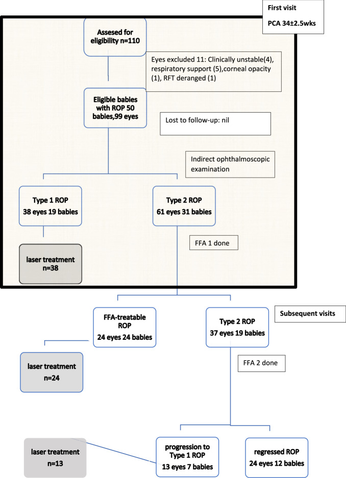
Subject/methods: Prospective single centre study in a tertiary care hospital of 99 eyes of 50 preterm babies. Fundus fluorescein angiography (FFA) was performed in all babies using RetCam 3 at the first detection of ROP. The babies were followed up for the progression of ROP. The FFA predictors for the progression of ROP were evaluated using the Mann-Whitney U test and Fisher's test.

Results: Thirty-eight eyes were Type 1 ROP at initial presentation and were lasered. Amongst the rest, 24 eyes showed features of stage 3 ROP with intense leakage on FFA and were designated as FFA-treatable ROP and were also lasered. Amongst the rest of the 37 eyes, the disease progression was seen in 13 eyes and the disease regression was seen in 24 eyes. The baseline FFA findings associated with the progression of ROP were delayed retinal arterial perfusion (p = 0.037) and popcorn lesions (p = 0.042). The post hoc analysis was done using a validated FFA scoring system.

Conclusions: FFA may be added in the classification of ROP and delayed retinal arterial perfusion and popcorn lesions on FFA may predict the progression of ROP.


Conflict of interest statement

The authors declare no competing interests.

Figures

Fig. 1
Fig. 1. Flow diagram of the study.
The shaded area on top represents the baby’s first visit for ROP screening to the Ophthalmologist.
Fig. 2
Fig. 2. FFA of a preterm baby with gestational age of 30 weeks and birth weight of 1000 g with stage I ROP.
A Arterio-venous phase showing preplus disease and popcorns (red plus sign), capillary non-perfusion areas (orange arrows), arterio-venous shunt vessels (yellow arrow heads), capillary tufts (pink ovoids) and rosary beads (green circle). B FFA late phase of the same baby 2 weeks later showing leakage from the ridge area and progression of plus disease.
Fig. 3
Fig. 3. Fundus angiography picture showing abnormal findings in ROP babies.
A FFA showing delayed arterio-venous filling at 1 min 40 s. The arrow demonstrates the unfilled retinal veins. B FFA arterio-venous phase showing avascular island superiorly lined by arterio-venous shunt vessels (Block black arrow). C FFA arterio-venous phase showing saccular dilatations of arterioles (seen in the white circle). D FFA arterio-venous phase showing arterio- venous shunt vessels in vascularized retina (black arrow head) and macular dye leakage suggestive of macular oedema (black arrow).

References

    1. Temkar S, Azad SV, Chawla R, Damodaran S, Garg G, Regani H, et al. Ultra-widefield fundus fluorescein angiography in pediatric retinal vascular diseases. Indian J Ophthal. 2019;67:788–94. doi: 10.4103/ijo.IJO_1688_18. - DOI - PMC - PubMed
    1. Early Treatment for Retinopathy of Prematurity Cooperative Group. Revised indications for the treatment of retinopathy of prematurity: results of the early treatment for retinopathy of prematurity randomized trial. Arch Ophthalmol. 2003;121:1684–94. doi: 10.1001/archopht.121.12.1684. - DOI - PubMed
    1. International Committee for Classification of Retinopathy of Prematurity. The international classification of retinopathy of prematurity revisited. Arch Ophthalmol. 2005;123:991–9. doi: 10.1001/archopht.123.7.991. - DOI - PubMed
    1. Christiansen SP, Dobson V, Quinn GE, Good WV, Tung B, Hardy RJ, et al. Progression of type 2 to type 1 retinopathy of prematurity in the Early Treatment for retinopathy of prematurity study. Arch Ophthalmol. 2010;128:461–5. doi: 10.1001/archophthalmol.2010.34. - DOI - PubMed
    1. Lepore D, Molle F, Pagliara MM, Baldascino A, Angora C, Sammartino M, et al. Atlas of fluorescein angiographic findings in eyes undergoing laser for retinopathy of prematurity. Ophthalmology. 2011;118:168–75. doi: 10.1016/j.ophtha.2010.04.021. - DOI - PubMed