

ST6GAL1 and α2-6 Sialylation Regulates IL-6 Expression and Secretion in Chronic Obstructive Pulmonary Disease
- PMID: 34290711
- PMCID: PMC8287524
- DOI: 10.3389/fimmu.2021.693149
ST6GAL1 and α2-6 Sialylation Regulates IL-6 Expression and Secretion in Chronic Obstructive Pulmonary Disease
Abstract
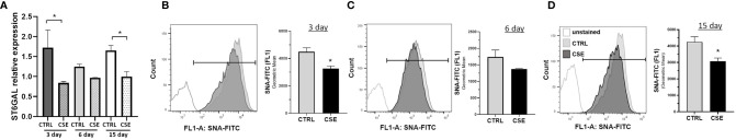
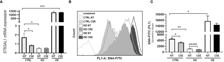
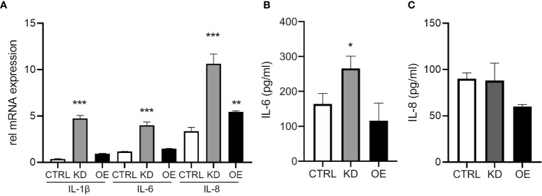
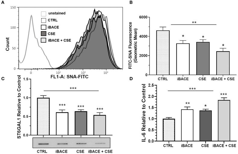
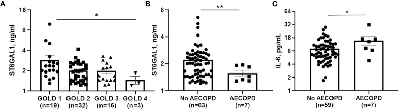
Chronic obstructive pulmonary disease (COPD) is a systemic disease strongly associated with cigarette smoking, airway inflammation, and acute disease exacerbations. Changes in terminal sialylation and fucosylation of asparagine (N)-linked glycans have been documented in COPD, but the role that glycosyltransferases may play in the regulation of N-linked glycans in COPD has not been fully elucidated. Recent studies suggest that modulation of ST6GAL1 (ST6 beta-galactoside alpha-2,6-sialyltransferase-1), which catalyzes terminal α2-6 sialylation of cellular proteins, may regulate inflammation and contribute to COPD phenotype(s). Interestingly, it has been previously demonstrated that ST6GAL1, a Golgi resident protein, can be proteolytically processed by BACE1 (beta-site amyloid precursor protein cleaving enzyme-1) to a circulating form that retains activity. In this study, we showed that loss of ST6GAL1 expression increased interleukin (IL)-6 expression and secretion in human bronchial epithelial cells (HBECs). Furthermore, exposure to cigarette smoke medium/extract (CSE) or BACE1 inhibition resulted in decreased ST6GAL1 secretion, reduced α2-6 sialylation, and increased IL-6 production in HBECs. Analysis of plasma ST6GAL1 levels in a small COPD patient cohort demonstrated an inverse association with prospective acute exacerbations of COPD (AECOPD), while IL-6 was positively associated. Altogether, these results suggest that reduced ST6GAL1 and α2-6 sialylation augments IL-6 expression/secretion in HBECs and is associated with poor clinical outcomes in COPD.
Keywords: COPD; ST6GAL1; bronchial epithelium; cigarette smoke; inflammation.
Copyright © 2021 Krick, Helton, Easter, Bollenbecker, Denson, Zaharias, Cochran, Vang, Harris, Wells and Barnes.
Conflict of interest statement
JW receives grant support from Bayer AG, Vertex Pharmaceuticals, Grifols, Verona Pharma, Mereo BioPharma, and Arcus-MED to conduct research independent from the work in the current manuscript and has been a consultant to AstraZeneca, Takeda, and GlaxoSmithKline for projects unrelated to the content of this work. The remaining authors declare that the research was conducted in the absence of any commercial or financial relationships that could be construed as a potential conflict of interest.
Figures

Similar articles
-
Sialylation of MUC4β N-glycans by ST6GAL1 orchestrates human airway epithelial cell differentiation associated with type-2 inflammation.JCI Insight. 2019 Mar 7;4(5):e122475. doi: 10.1172/jci.insight.122475. eCollection 2019 Mar 7. JCI Insight. 2019. PMID: 30730306 Free PMC article.
-
Loss of core fucosylation in both ST6GAL1 and its substrate enhances glycoprotein sialylation in mice.Biochem J. 2020 Mar 27;477(6):1179-1201. doi: 10.1042/BCJ20190789. Biochem J. 2020. PMID: 32141499
-
β-Galactoside α2,6-sialyltranferase 1 promotes transforming growth factor-β-mediated epithelial-mesenchymal transition.J Biol Chem. 2014 Dec 12;289(50):34627-41. doi: 10.1074/jbc.M114.593392. Epub 2014 Oct 24. J Biol Chem. 2014. PMID: 25344606 Free PMC article.
-
Specific sialylation of N-glycans and its novel regulatory mechanism.Glycoconj J. 2024 Jun;41(3):175-183. doi: 10.1007/s10719-024-10157-8. Epub 2024 Jul 3. Glycoconj J. 2024. PMID: 38958800 Free PMC article. Review.
-
Regulation of ST6GAL1 sialyltransferase expression in cancer cells.Glycobiology. 2021 Jun 3;31(5):530-539. doi: 10.1093/glycob/cwaa110. Glycobiology. 2021. PMID: 33320246 Free PMC article. Review.
Cited by
-
Targeting Non-Eosinophilic Immunological Pathways in COPD and AECOPD: Current Insights and Therapeutic Strategies.Int J Chron Obstruct Pulmon Dis. 2025 Mar 5;20:511-532. doi: 10.2147/COPD.S506616. eCollection 2025. Int J Chron Obstruct Pulmon Dis. 2025. PMID: 40066199 Free PMC article. Review.
-
Comparative electron microscopic visualization of the lung alveolar epithelial glycocalyx with different staining and labeling methods.J Anat. 2025 May;246(5):770-781. doi: 10.1111/joa.14129. Epub 2024 Sep 8. J Anat. 2025. PMID: 39245632 Free PMC article.
-
Synovial Fibroblast Sialylation Regulates Cell Migration and Activation of Inflammatory Pathways in Arthritogenesis.Front Immunol. 2022 Mar 18;13:847581. doi: 10.3389/fimmu.2022.847581. eCollection 2022. Front Immunol. 2022. PMID: 35371069 Free PMC article.
-
In vitro evidence of antioxidant and anti-inflammatory effects of a new nutraceutical formulation explains benefits in a clinical setting of COPD patients.Front Pharmacol. 2024 Aug 20;15:1439835. doi: 10.3389/fphar.2024.1439835. eCollection 2024. Front Pharmacol. 2024. PMID: 39228520 Free PMC article.
-
ST6Gal1: Oncogenic signaling pathways and targets.Front Mol Biosci. 2022 Aug 29;9:962908. doi: 10.3389/fmolb.2022.962908. eCollection 2022. Front Mol Biosci. 2022. PMID: 36106023 Free PMC article. Review.
References
-
- Varki A, Cummings RD, Esko JD, Freeze HH, Stanley P, Bertozzi CR, et al. . Essential. In: Glycobiology. Cold Spring Harbor. NY: Cold Spring Harbor Laboratory Press; (2009). - PubMed
Publication types
MeSH terms
Substances
Grants and funding
LinkOut - more resources
Full Text Sources
Medical